 Movie
Movie Controller


+ Open data

- Basic information
| Entry | Database: PDB / ID: 5gl1 | |||||||||
|---|---|---|---|---|---|---|---|---|---|---|
| Title | Structure of RyR1 in an open state | |||||||||
 Components Components |
| |||||||||
 Keywords Keywords | TRANSPORT PROTEIN/ISOMERASE / channel / membrane protein / TRANSPORT PROTEIN-ISOMERASE complex | |||||||||
| Function / homology |  Function and homology information Function and homology informationcytoplasmic side of membrane / ATP-gated ion channel activity / ryanodine-sensitive calcium-release channel activity / terminal cisterna / ryanodine receptor complex / release of sequestered calcium ion into cytosol by sarcoplasmic reticulum / ossification involved in bone maturation / cellular response to caffeine / skin development / organelle membrane ...cytoplasmic side of membrane / ATP-gated ion channel activity / ryanodine-sensitive calcium-release channel activity / terminal cisterna / ryanodine receptor complex / release of sequestered calcium ion into cytosol by sarcoplasmic reticulum / ossification involved in bone maturation / cellular response to caffeine / skin development / organelle membrane / smooth endoplasmic reticulum / outflow tract morphogenesis / intracellularly gated calcium channel activity / intracellular membrane-bounded organelle / regulation of ryanodine-sensitive calcium-release channel activity / toxic substance binding / voltage-gated calcium channel activity / skeletal muscle fiber development / regulation of release of sequestered calcium ion into cytosol by sarcoplasmic reticulum / release of sequestered calcium ion into cytosol / muscle contraction / sarcoplasmic reticulum membrane / striated muscle contraction / cellular response to calcium ion / peptidylprolyl isomerase / sarcoplasmic reticulum / peptidyl-prolyl cis-trans isomerase activity / sarcolemma / intracellular calcium ion homeostasis / Z disc / calcium channel activity / calcium ion transmembrane transport / disordered domain specific binding / protein homotetramerization / transmembrane transporter binding / calmodulin binding / calcium ion binding / ATP binding / membrane / identical protein binding / cytosol Similarity search - Function | |||||||||
| Biological species |  | |||||||||
| Method | ELECTRON MICROSCOPY / single particle reconstruction / cryo EM / Resolution: 5.7 Å | |||||||||
 Authors Authors | Bai, X.C. / Yan, Z. / Wu, J.P. / Yan, N. | |||||||||
| Funding support |  China, 2items China, 2items
| |||||||||
 Citation Citation | Journal: Cell Res / Year: 2016 Title: The Central domain of RyR1 is the transducer for long-range allosteric gating of channel opening. Authors: Xiao-Chen Bai / Zhen Yan / Jianping Wu / Zhangqiang Li / Nieng Yan /  Abstract: The ryanodine receptors (RyRs) are intracellular calcium channels responsible for rapid release of Ca(2+) from the sarcoplasmic/endoplasmic reticulum (SR/ER) to the cytoplasm, which is essential for ...The ryanodine receptors (RyRs) are intracellular calcium channels responsible for rapid release of Ca(2+) from the sarcoplasmic/endoplasmic reticulum (SR/ER) to the cytoplasm, which is essential for the excitation-contraction (E-C) coupling of cardiac and skeletal muscles. The near-atomic resolution structure of closed RyR1 revealed the molecular details of this colossal channel, while the long-range allosteric gating mechanism awaits elucidation. Here, we report the cryo-EM structures of rabbit RyR1 in three closed conformations at about 4 Å resolution and an open state at 5.7 Å. Comparison of the closed RyR1 structures shows a breathing motion of the cytoplasmic platform, while the channel domain and its contiguous Central domain remain nearly unchanged. Comparison of the open and closed structures shows a dilation of the S6 tetrahelical bundle at the cytoplasmic gate that leads to channel opening. During the pore opening, the cytoplasmic "O-ring" motif of the channel domain and the U-motif of the Central domain exhibit coupled motion, while the Central domain undergoes domain-wise displacement. These structural analyses provide important insight into the E-C coupling in skeletal muscles and identify the Central domain as the transducer that couples the conformational changes of the cytoplasmic platform to the gating of the central pore. | |||||||||
| History |
|
- Structure visualization
| Movie |
Movie viewer |
|---|---|
| Structure viewer | Molecule: MolmilJmol/JSmol |
- Downloads & links
-Download
| PDBx/mmCIF format | 5gl1.cif.gz | 2.7 MB | Display | PDBx/mmCIF format |
|---|---|---|---|---|
| PDB format | pdb5gl1.ent.gz | Display | PDB format | |
| PDBx/mmJSON format | 5gl1.json.gz | Tree view | PDBx/mmJSON format | |
| Others |  Other downloads Other downloads |
-Validation report
| Arichive directory | https://data.pdbj.org/pub/pdb/validation_reports/gl/5gl1ftp://data.pdbj.org/pub/pdb/validation_reports/gl/5gl1 | HTTPS FTP |
|---|
-Related structure data
| Related structure data | 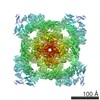 9521MC 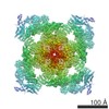 9518C 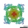 9519C 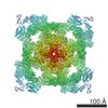 9520C  5gkyC  5gkzC  5gl0C M: map data used to model this data C: citing same article ( |
|---|---|
| Similar structure data |


-Links
 PDBj
PDBj

- Assembly
| Deposited unit | 
|
|---|---|
| 1 |
|
-Components
| #1: Protein | Mass: 565908.625 Da / Num. of mol.: 4 / Source method: isolated from a natural source / Source: (natural) #2: Protein | Mass: 11967.705 Da / Num. of mol.: 4 Source method: isolated from a genetically manipulated source Source: (gene. exp.)  #3: Chemical | ChemComp-ZN /   Mass: 65.409 Da / Num. of mol.: 4 / Source method: obtained synthetically / Formula: Zn Mass: 65.409 Da / Num. of mol.: 4 / Source method: obtained synthetically / Formula: Zn |
|---|
-Experimental details
-Experiment
| Experiment | Method: ELECTRON MICROSCOPY |
|---|---|
| EM experiment | Aggregation state: PARTICLE / 3D reconstruction method: single particle reconstruction |
- Sample preparation
| Component |
| ||||||||||||||||||||||||
|---|---|---|---|---|---|---|---|---|---|---|---|---|---|---|---|---|---|---|---|---|---|---|---|---|---|
| Molecular weight | Value: 2.2 MDa / Experimental value: NO | ||||||||||||||||||||||||
| Source (natural) |
| ||||||||||||||||||||||||
| Source (recombinant) | Organism: | ||||||||||||||||||||||||
| Buffer solution | pH: 7.4 Details: 20 mM MOPS-Na, pH 7.4, 250 mM NaCl, 2 mM DTT, 0.5% CHAPS (w/v) (Sigma-Aldrich) and protease inhibitor cocktail including 2 mM PMSF, 2.6 ug/ml aprotinin, 1.4 ug/ml pepstatin, and 10 ug/ml ...Details: 20 mM MOPS-Na, pH 7.4, 250 mM NaCl, 2 mM DTT, 0.5% CHAPS (w/v) (Sigma-Aldrich) and protease inhibitor cocktail including 2 mM PMSF, 2.6 ug/ml aprotinin, 1.4 ug/ml pepstatin, and 10 ug/ml leupeptin (Amresco),50 uM CaCl2 and 10 uM PCB95 | ||||||||||||||||||||||||
| Specimen | Conc.: 0.066 mg/ml / Embedding applied: NO / Shadowing applied: NO / Staining applied: NO / Vitrification applied: YES | ||||||||||||||||||||||||
| Specimen support | Grid material: COPPER / Grid mesh size: 200 divisions/in. / Grid type: Quantifoil R2/2 | ||||||||||||||||||||||||
| Vitrification | Cryogen name: ETHANE / Humidity: 100 % / Chamber temperature: 277 K |
- Electron microscopy imaging
| Experimental equipment |  Model: Tecnai Polara / Image courtesy: FEI Company |
|---|---|
| Microscopy | Model: FEI POLARA 300 |
| Electron gun | Electron source:  FIELD EMISSION GUN / Accelerating voltage: 300 kV / Illumination mode: FLOOD BEAM FIELD EMISSION GUN / Accelerating voltage: 300 kV / Illumination mode: FLOOD BEAM |
| Electron lens | Mode: BRIGHT FIELD |
| Specimen holder | Cryogen: NITROGEN |
| Image recording | Electron dose: 40 e/Å2 / Film or detector model: FEI FALCON II (4k x 4k) |
- Processing
| EM software |
| ||||||||||||||||||||||||||||
|---|---|---|---|---|---|---|---|---|---|---|---|---|---|---|---|---|---|---|---|---|---|---|---|---|---|---|---|---|---|
| Image processing | Details: Actually we use a prototype FEI Falcon-III detector | ||||||||||||||||||||||||||||
| CTF correction | Type: PHASE FLIPPING AND AMPLITUDE CORRECTION | ||||||||||||||||||||||||||||
| 3D reconstruction | Resolution: 5.7 Å / Resolution method: FSC 0.143 CUT-OFF / Num. of particles: 30000 / Symmetry type: POINT |
