 Movie
Movie Controller

[English] 日本語
 Yorodumi
- PDB-5a21: Structure of bacteriophage SPP1 head-to-tail interface without DN... -
+ Open data

- Basic information
| Entry | Database: PDB / ID: 5a21 | ||||||
|---|---|---|---|---|---|---|---|
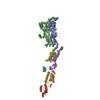
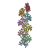
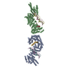
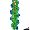
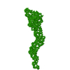
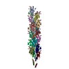
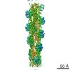
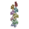
| Title | Structure of bacteriophage SPP1 head-to-tail interface without DNA and tape measure protein | ||||||
 Components Components |
| ||||||
 Keywords Keywords | VIRAL PROTEIN / VIRAL INFECTION / TAILED BACTERIOPHAGE / SIPHOVIRIDAE / SPP1 / VIRAL ASSEMBLY / HEAD-TO-TAIL INTERFACE / DNA GATEKEEPER / ALLOSTERIC MECHANISM / CONCERTED REORGANISATION / DIAPHRAGM GATING | ||||||
| Function / homology |  Function and homology information Function and homology informationviral head-tail joining / virus tail fiber assembly / viral DNA genome packaging, headful / virus tail, tube / viral procapsid / viral portal complex / symbiont genome ejection through host cell envelope, long flexible tail mechanism / virus tail / virion component / viral translational frameshifting Similarity search - Function | ||||||
| Biological species |  BACILLUS PHAGE SPP1 (virus) BACILLUS PHAGE SPP1 (virus) | ||||||
| Method | ELECTRON MICROSCOPY / single particle reconstruction / cryo EM / Resolution: 7.2 Å | ||||||
 Authors Authors | Chaban, Y. / Lurz, R. / Brasiles, S. / Cornilleau, C. / Karreman, M. / Zinn-Justin, S. / Tavares, P. / Orlova, E.V. | ||||||
 Citation Citation | Journal: Proc Natl Acad Sci U S A / Year: 2015 Title: Structural rearrangements in the phage head-to-tail interface during assembly and infection. Authors: Yuriy Chaban / Rudi Lurz / Sandrine Brasilès / Charlène Cornilleau / Matthia Karreman / Sophie Zinn-Justin / Paulo Tavares / Elena V Orlova /    Abstract: Many icosahedral viruses use a specialized portal vertex to control genome encapsidation and release from the viral capsid. In tailed bacteriophages, the portal system is connected to a tail ...Many icosahedral viruses use a specialized portal vertex to control genome encapsidation and release from the viral capsid. In tailed bacteriophages, the portal system is connected to a tail structure that provides the pipeline for genome delivery to the host cell. We report the first, to our knowledge, subnanometer structures of the complete portal-phage tail interface that mimic the states before and after DNA release during phage infection. They uncover structural rearrangements associated with intimate protein-DNA interactions. The portal protein gp6 of bacteriophage SPP1 undergoes a concerted reorganization of the structural elements of its central channel during interaction with DNA. A network of protein-protein interactions primes consecutive binding of proteins gp15 and gp16 to extend and close the channel. This critical step that prevents genome leakage from the capsid is achieved by a previously unidentified allosteric mechanism: gp16 binding to two different regions of gp15 drives correct positioning and folding of an inner gp16 loop to interact with equivalent loops of the other gp16 subunits. Together, these loops build a plug that closes the channel. Gp16 then fastens the tail to yield the infectious virion. The gatekeeper system opens for viral genome exit at the beginning of infection but recloses afterward, suggesting a molecular diaphragm-like mechanism to control DNA efflux. The mechanisms described here, controlling the essential steps of phage genome movements during virus assembly and infection, are likely to be conserved among long-tailed phages, the largest group of viruses in the Biosphere. | ||||||
| History |
|
- Structure visualization
| Movie |
Movie viewer |
|---|---|
| Structure viewer | Molecule: MolmilJmol/JSmol |
- Downloads & links
-Download
| PDBx/mmCIF format | 5a21.cif.gz | 296.9 KB | Display | PDBx/mmCIF format |
|---|---|---|---|---|
| PDB format | pdb5a21.ent.gz | 233.3 KB | Display | PDB format |
| PDBx/mmJSON format | 5a21.json.gz | Tree view | PDBx/mmJSON format | |
| Others |  Other downloads Other downloads |
-Validation report
| Arichive directory | https://data.pdbj.org/pub/pdb/validation_reports/a2/5a21ftp://data.pdbj.org/pub/pdb/validation_reports/a2/5a21 | HTTPS FTP |
|---|
-Related structure data
| Related structure data | 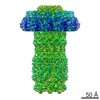 2994MC 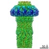 2993C 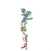 5a20C C: citing same article ( M: map data used to model this data |
|---|---|
| Similar structure data |
-Links
 PDBj
PDBj


- Assembly
| Deposited unit | 
|
|---|---|
| 1 |
|
-Components
| #1: Protein | Mass: 57405.355 Da / Num. of mol.: 2 / Source method: isolated from a natural source / Source: (natural) BACILLUS PHAGE SPP1 (virus) / References: UniProt: P54309#2: Protein | Mass: 11629.402 Da / Num. of mol.: 2 / Source method: isolated from a natural source / Source: (natural) BACILLUS PHAGE SPP1 (virus) / References: UniProt: Q38584#3: Protein | Mass: 12615.069 Da / Num. of mol.: 2 / Source method: isolated from a natural source / Source: (natural) BACILLUS PHAGE SPP1 (virus) / References: UniProt: O48446#4: Protein | | Mass: 15025.014 Da / Num. of mol.: 1 / Source method: isolated from a natural source / Source: (natural) BACILLUS PHAGE SPP1 (virus) / References: UniProt: O48448#5: Protein | | Mass: 19172.906 Da / Num. of mol.: 1 / Source method: isolated from a natural source / Source: (natural) BACILLUS PHAGE SPP1 (virus) / References: UniProt: O48449 |
|---|
-Experimental details
-Experiment
| Experiment | Method: ELECTRON MICROSCOPY |
|---|---|
| EM experiment | Aggregation state: PARTICLE / 3D reconstruction method: single particle reconstruction |
- Sample preparation
| Component | Name: BACTERIOPHAGE SPP1 HEAD- TO-TAIL INTERFACE / Type: VIRUS / Details: MICROGRAPHS SELECTED BY OPTICAL DIFFRACTION |
|---|---|
| Buffer solution | Name: SEE REFERENCE FOR DETAILS / pH: 7.5 / Details: SEE REFERENCE FOR DETAILS |
| Specimen | Embedding applied: NO / Shadowing applied: NO / Staining applied: NO / Vitrification applied: YES |
| Specimen support | Details: CARBON |
| Vitrification | Instrument: FEI VITROBOT MARK II / Cryogen name: ETHANE Details: VITRIFICATION 1 -- CRYOGEN- ETHANE, INSTRUMENT- FEI VITROBOT |
- Electron microscopy imaging
| Experimental equipment |  Model: Tecnai F30 / Image courtesy: FEI Company |
|---|---|
| Microscopy | Model: FEI TECNAI F30 / Date: Oct 9, 2008 |
| Electron gun | Electron source:  FIELD EMISSION GUN / Accelerating voltage: 300 kV / Illumination mode: FLOOD BEAM FIELD EMISSION GUN / Accelerating voltage: 300 kV / Illumination mode: FLOOD BEAM |
| Electron lens | Mode: BRIGHT FIELD / Nominal magnification: 39000 X / Nominal defocus max: 3600 nm / Nominal defocus min: 900 nm / Cs: 2 mm |
| Image recording | Electron dose: 20 e/Å2 / Film or detector model: KODAK SO-163 FILM |
| Image scans | Num. digital images: 400 |
- Processing
| EM software |
| ||||||||||||||||||||||||||||||||
|---|---|---|---|---|---|---|---|---|---|---|---|---|---|---|---|---|---|---|---|---|---|---|---|---|---|---|---|---|---|---|---|---|---|
| CTF correction | Details: EACH PARTICLE | ||||||||||||||||||||||||||||||||
| Symmetry | Point symmetry: C6 (6 fold cyclic) | ||||||||||||||||||||||||||||||||
| 3D reconstruction | Method: ANGULAR RECONSTITUTION / Resolution: 7.2 Å / Num. of particles: 18000 / Nominal pixel size: 1.2 Å / Actual pixel size: 1.15 Å Magnification calibration: CROSS- -CORRELATION WITH FITTED ATOMIC COORDINATES Details: ATOMIC COORDINATES FOR GP6, GP15, GP16, GP17 WERE OBTAINED FROM PDB FILES 2JES (LEBEDEV ET AL., EMBO J., 2007, 26, 1984), 2KBZ, 2KCA (LHUILLIER ET AL., PROC.NATL.ACAD.SCI. USA, 2009, 106, ...Details: ATOMIC COORDINATES FOR GP6, GP15, GP16, GP17 WERE OBTAINED FROM PDB FILES 2JES (LEBEDEV ET AL., EMBO J., 2007, 26, 1984), 2KBZ, 2KCA (LHUILLIER ET AL., PROC.NATL.ACAD.SCI. USA, 2009, 106, 8507), 2LFP (CHAGOT ET AL., PROTEINS, 2012, 80, 319), CORRESPONDIGLY, AND DOCKED INTO EM ELECTRON DENSITY MAP USING FLEXIBLE FIT. ATOMIC COORDINATES FOR MISSING DOMAINS OF GP6 AND GP17.1. WERE MODELLED USING I-TASSER PROTEIN STRUCTURE PREDICTION SERVER (Y ZHANG, BMC BIOINFORMATICS, 2008, 9, 40) AND DOCKED INTO EM ELECTRON DENSITY MAP USING FLEXIBLE FIT. SUBMISSION BASED ON EXPERIMENTAL DATA FROM EMDB EMD-2994. (DEPOSITION ID: 13332). Symmetry type: POINT | ||||||||||||||||||||||||||||||||
| Atomic model building | Protocol: FLEXIBLE FIT / Space: REAL Details: METHOD--FLEXIBLE REFINEMENT PROTOCOL--X-RAY, NMR, PREDICTION | ||||||||||||||||||||||||||||||||
| Refinement | Highest resolution: 7.2 Å | ||||||||||||||||||||||||||||||||
| Refinement step | Cycle: LAST / Highest resolution: 7.2 Å
|
