 Movie
Movie Controller

[English] 日本語
 Yorodumi
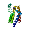
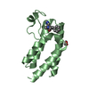
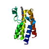
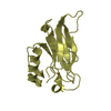
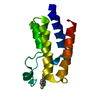
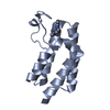
- PDB-5or8: Crystal Structure of BAZ2A bromodomain in complex with 1,3-dimeth... -
+ Open data

- Basic information
| Entry | Database: PDB / ID: 5or8 | ||||||
|---|---|---|---|---|---|---|---|
| Title | Crystal Structure of BAZ2A bromodomain in complex with 1,3-dimethyl-benzimidazolone compound 1 | ||||||
 Components Components | Bromodomain adjacent to zinc finger domain protein 2A | ||||||
 Keywords Keywords | TRANSCRIPTION / four helical bundle | ||||||
| Function / homology |  Function and homology information Function and homology informationNoRC complex / rDNA heterochromatin / rDNA heterochromatin formation / RNA polymerase I preinitiation complex assembly / chromatin silencing complex / negative regulation of transcription by RNA polymerase I / DNA methylation-dependent constitutive heterochromatin formation / histone H4K16ac reader activity / nuclear receptor binding / NoRC negatively regulates rRNA expression ...NoRC complex / rDNA heterochromatin / rDNA heterochromatin formation / RNA polymerase I preinitiation complex assembly / chromatin silencing complex / negative regulation of transcription by RNA polymerase I / DNA methylation-dependent constitutive heterochromatin formation / histone H4K16ac reader activity / nuclear receptor binding / NoRC negatively regulates rRNA expression / heterochromatin formation / histone binding / nuclear speck / chromatin remodeling / regulation of DNA-templated transcription / DNA-templated transcription / nucleolus / DNA binding / RNA binding / zinc ion binding / nucleus / cytosol Similarity search - Function | ||||||
| Biological species |  Homo sapiens (human) Homo sapiens (human) | ||||||
| Method |  X-RAY DIFFRACTION / SYNCHROTRON / MOLECULAR REPLACEMENT / molecular replacement / Resolution: 2.4 Å X-RAY DIFFRACTION / SYNCHROTRON / MOLECULAR REPLACEMENT / molecular replacement / Resolution: 2.4 Å | ||||||
 Authors Authors | Lolli, G. / Dalle Vedove, A. / Marchand, J.-R. / Caflisch, A. | ||||||
| Funding support |  Switzerland, 1items Switzerland, 1items
| ||||||
 Citation Citation | Journal: J Chem Inf Model / Year: 2017 Title: Discovery of Inhibitors of Four Bromodomains by Fragment-Anchored Ligand Docking. Authors: Marchand, J.R. / Dalle Vedove, A. / Lolli, G. / Caflisch, A. | ||||||
| History |
|
- Structure visualization
| Structure viewer | Molecule: MolmilJmol/JSmol |
|---|
- Downloads & links
-Download
| PDBx/mmCIF format | 5or8.cif.gz | 36.9 KB | Display | PDBx/mmCIF format |
|---|---|---|---|---|
| PDB format | pdb5or8.ent.gz | 23.5 KB | Display | PDB format |
| PDBx/mmJSON format | 5or8.json.gz | Tree view | PDBx/mmJSON format | |
| Others |  Other downloads Other downloads |
-Validation report
| Arichive directory | https://data.pdbj.org/pub/pdb/validation_reports/or/5or8ftp://data.pdbj.org/pub/pdb/validation_reports/or/5or8 | HTTPS FTP |
|---|
-Related structure data
| Related structure data | 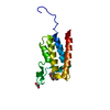 5or9C  5orbC 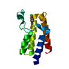 5mgjS S: Starting model for refinement C: citing same article ( |
|---|---|
| Similar structure data |


-Links
 PDBj
PDBj


- Assembly
| Deposited unit | 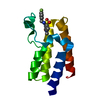
| ||||||||
|---|---|---|---|---|---|---|---|---|---|
| 1 |
| ||||||||
| Unit cell |
|
-Components
| #1: Protein | Mass: 12509.048 Da / Num. of mol.: 1 / Fragment: Bromodomain, UNP residues 1796-1899 Mutation: First two residues SM derive from the expression tag Source method: isolated from a genetically manipulated source Details: First two residues SM derive from the expression tag Source: (gene. exp.) Homo sapiens (human) / Gene: BAZ2A, KIAA0314, TIP5 / Production host:  |
|---|---|
| #2: Chemical | ChemComp-JR4 / ~{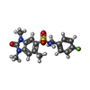  Mass: 363.407 Da / Num. of mol.: 1 / Source method: obtained synthetically / Formula: C17H18FN3O3S Mass: 363.407 Da / Num. of mol.: 1 / Source method: obtained synthetically / Formula: C17H18FN3O3S |
| #3: Water | ChemComp-HOH /  Mass: 18.015 Da / Num. of mol.: 49 / Source method: isolated from a natural source / Formula: H2O Mass: 18.015 Da / Num. of mol.: 49 / Source method: isolated from a natural source / Formula: H2O |
-Experimental details
-Experiment
| Experiment | Method: X-RAY DIFFRACTION / Number of used crystals: 1 |
|---|
- Sample preparation
| Crystal | Density Matthews: 3.51 Å3/Da / Density % sol: 64.93 % / Mosaicity: 0.2 ° |
|---|---|
| Crystal grow | Temperature: 277 K / Method: vapor diffusion, sitting drop / pH: 7.5 / Details: 20% PEG3350, 0.2 M MgCl2 |
-Data collection
| Diffraction | Mean temperature: 100 K | ||||||||||||||||||||||||
|---|---|---|---|---|---|---|---|---|---|---|---|---|---|---|---|---|---|---|---|---|---|---|---|---|---|
| Diffraction source | Source: SYNCHROTRON / Site: ELETTRA  / Beamline: 5.2R / Wavelength: 1 Å / Beamline: 5.2R / Wavelength: 1 Å | ||||||||||||||||||||||||
| Detector | Type: DECTRIS PILATUS 2M / Detector: PIXEL / Date: Jul 5, 2017 | ||||||||||||||||||||||||
| Radiation | Protocol: SINGLE WAVELENGTH / Monochromatic (M) / Laue (L): M / Scattering type: x-ray | ||||||||||||||||||||||||
| Radiation wavelength | Wavelength: 1 Å / Relative weight: 1 | ||||||||||||||||||||||||
| Reflection | Resolution: 2.4→47.57 Å / Num. obs: 6827 / % possible obs: 99.9 % / Redundancy: 9.9 % / CC1/2: 0.991 / Rmerge(I) obs: 0.258 / Rpim(I) all: 0.086 / Rrim(I) all: 0.272 / Net I/σ(I): 8.3 | ||||||||||||||||||||||||
| Reflection shell | Diffraction-ID: 1
|
-Phasing
| Phasing | Method: molecular replacement |
|---|
- Processing
| Software |
| ||||||||||||||||||||||||
|---|---|---|---|---|---|---|---|---|---|---|---|---|---|---|---|---|---|---|---|---|---|---|---|---|---|
| Refinement | Method to determine structure: MOLECULAR REPLACEMENT Starting model: 5MGJ Resolution: 2.4→41.195 Å / SU ML: 0.24 / Cross valid method: FREE R-VALUE / σ(F): 1.34 / Phase error: 23.17
| ||||||||||||||||||||||||
| Solvent computation | Shrinkage radii: 0.9 Å / VDW probe radii: 1.11 Å | ||||||||||||||||||||||||
| Displacement parameters | Biso max: 92.06 Å2 / Biso mean: 40.223 Å2 / Biso min: 19.02 Å2 | ||||||||||||||||||||||||
| Refinement step | Cycle: final / Resolution: 2.4→41.195 Å
| ||||||||||||||||||||||||
| Refine LS restraints |
| ||||||||||||||||||||||||
| LS refinement shell | Refine-ID: X-RAY DIFFRACTION / Rfactor Rfree error: 0 / Total num. of bins used: 2 / % reflection obs: 100 %
|
