 Movie
Movie Controller

[English] 日本語
 Yorodumi
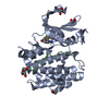
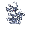
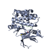
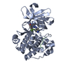
- PDB-5n5l: Crystal structure of human Pim-1 kinase in complex with a consens... -
+ Open data

- Basic information
| Entry | Database: PDB / ID: 5n5l | ||||||
|---|---|---|---|---|---|---|---|
| Title | Crystal structure of human Pim-1 kinase in complex with a consensuspeptide and [2-oxo-2-(1H-pyrrol-2-yl)ethyl] 5-bromo-1H-indole-3-carboxylate | ||||||
 Components Components |
| ||||||
 Keywords Keywords | TRANSFERASE / Serine Threonine Kinase / proto-oncogene / Fragment / PIM-1 / Consensus peptide | ||||||
| Function / homology |  Function and homology information Function and homology informationregulation of transmembrane transporter activity / positive regulation of cardioblast proliferation / positive regulation of cyclin-dependent protein serine/threonine kinase activity / regulation of hematopoietic stem cell proliferation / vitamin D receptor signaling pathway / cellular detoxification / STAT5 activation downstream of FLT3 ITD mutants / transcription factor binding / positive regulation of protein serine/threonine kinase activity / ribosomal small subunit binding ...regulation of transmembrane transporter activity / positive regulation of cardioblast proliferation / positive regulation of cyclin-dependent protein serine/threonine kinase activity / regulation of hematopoietic stem cell proliferation / vitamin D receptor signaling pathway / cellular detoxification / STAT5 activation downstream of FLT3 ITD mutants / transcription factor binding / positive regulation of protein serine/threonine kinase activity / ribosomal small subunit binding / : / positive regulation of cardiac muscle cell proliferation / positive regulation of brown fat cell differentiation / Signaling by FLT3 fusion proteins / positive regulation of TORC1 signaling / regulation of mitotic cell cycle / negative regulation of innate immune response / protein serine/threonine kinase activator activity / cellular response to type II interferon / protein autophosphorylation / manganese ion binding / Interleukin-4 and Interleukin-13 signaling / protein phosphorylation / non-specific serine/threonine protein kinase / protein stabilization / protein serine kinase activity / protein serine/threonine kinase activity / apoptotic process / negative regulation of apoptotic process / positive regulation of DNA-templated transcription / nucleolus / nucleoplasm / ATP binding / nucleus / plasma membrane / cytoplasm / cytosol Similarity search - Function | ||||||
| Biological species |  Homo sapiens (human) Homo sapiens (human) | ||||||
| Method |  X-RAY DIFFRACTION / SYNCHROTRON / MOLECULAR REPLACEMENT / Resolution: 1.97 Å X-RAY DIFFRACTION / SYNCHROTRON / MOLECULAR REPLACEMENT / Resolution: 1.97 Å | ||||||
 Authors Authors | Siefker, C. / Heine, A. / Klebe, G. | ||||||
 Citation Citation | Journal: to be published Title: A crystallographic fragment study with human Pim-1 kinase Authors: Siefker, C. / Heine, A. / Taylor, C. / Kolb, P. / Hardes, K. / Steinmetzer, A. / Klebe, G. | ||||||
| History |
|
- Structure visualization
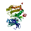
| Structure viewer | Molecule: MolmilJmol/JSmol |
|---|
- Downloads & links
-Download
| PDBx/mmCIF format | 5n5l.cif.gz | 133.2 KB | Display | PDBx/mmCIF format |
|---|---|---|---|---|
| PDB format | pdb5n5l.ent.gz | 103.5 KB | Display | PDB format |
| PDBx/mmJSON format | 5n5l.json.gz | Tree view | PDBx/mmJSON format | |
| Others |  Other downloads Other downloads |
-Validation report
| Arichive directory | https://data.pdbj.org/pub/pdb/validation_reports/n5/5n5lftp://data.pdbj.org/pub/pdb/validation_reports/n5/5n5l | HTTPS FTP |
|---|
-Related structure data
| Related structure data | 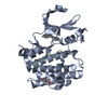 5mzlC 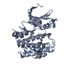 5n4nC 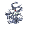 5n4oC 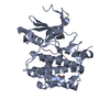 5n4rC  5n4uC 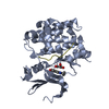 5n4vC 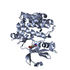 5n4xC  5n4yC 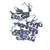 5n4zC 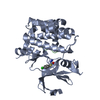 5n50C 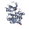 5n51C  5n52C 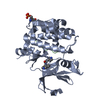 5n5mC 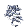 5ndtC  3we8S S: Starting model for refinement C: citing same article ( |
|---|---|
| Similar structure data |


-Links
 PDBj
PDBj

- Assembly
| Deposited unit | 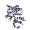
| ||||||||
|---|---|---|---|---|---|---|---|---|---|
| 1 |
| ||||||||
| Unit cell |
|
-Components
| #1: Protein | Mass: 35712.578 Da / Num. of mol.: 1 / Mutation: R250G Source method: isolated from a genetically manipulated source Details: Isofrom 2 of PIM-1 kinase Phosphorylation of Ser261 Source: (gene. exp.) Homo sapiens (human) / Gene: PIM1 / Plasmid: pLIC-SGC / Production host:  References: UniProt: P11309, non-specific serine/threonine protein kinase |
|---|---|
| #2: Protein/peptide | Mass: 1592.850 Da / Num. of mol.: 1 / Source method: obtained synthetically / Details: PIM-1 consensus peptide / Source: (synth.) Homo sapiens (human) |
| #3: Chemical | ChemComp-8NZ /   Mass: 240.053 Da / Num. of mol.: 1 / Source method: obtained synthetically / Formula: C9H6BrNO2 Mass: 240.053 Da / Num. of mol.: 1 / Source method: obtained synthetically / Formula: C9H6BrNO2 |
| #4: Chemical | ChemComp-GOL /   Mass: 92.094 Da / Num. of mol.: 1 / Source method: obtained synthetically / Formula: C3H8O3 Mass: 92.094 Da / Num. of mol.: 1 / Source method: obtained synthetically / Formula: C3H8O3 |
| #5: Water | ChemComp-HOH /  Mass: 18.015 Da / Num. of mol.: 166 / Source method: isolated from a natural source / Formula: H2O Mass: 18.015 Da / Num. of mol.: 166 / Source method: isolated from a natural source / Formula: H2O |
| Has protein modification | Y |
-Experimental details
-Experiment
| Experiment | Method: X-RAY DIFFRACTION / Number of used crystals: 1 |
|---|
- Sample preparation
| Crystal | Density Matthews: 3.1 Å3/Da / Density % sol: 60 % / Description: hexagonal rod |
|---|---|
| Crystal grow | Temperature: 277 K / Method: vapor diffusion, sitting drop / pH: 7 Details: HEPES Li2SO4 Glycerol DTT BIS-TRIS-propane PEG3350 Ethylene-glycol DMSO |
-Data collection
| Diffraction | Mean temperature: 100 K |
|---|---|
| Diffraction source | Source: SYNCHROTRON / Site: BESSY  / Beamline: 14.2 / Wavelength: 0.918409 Å / Beamline: 14.2 / Wavelength: 0.918409 Å |
| Detector | Type: DECTRIS PILATUS3 2M / Detector: PIXEL / Date: Sep 16, 2016 / Details: Silicon |
| Radiation | Monochromator: Si-111 crystal monochromator / Protocol: SINGLE WAVELENGTH / Monochromatic (M) / Laue (L): M / Scattering type: x-ray |
| Radiation wavelength | Wavelength: 0.918409 Å / Relative weight: 1 |
| Reflection | Resolution: 1.97→50 Å / Num. obs: 30857 / % possible obs: 99.9 % / Redundancy: 6.84 % / CC1/2: 0.99 / Rrim(I) all: 0.076 / Net I/σ(I): 16.63 |
| Reflection shell | Resolution: 1.97→2.09 Å / Redundancy: 6.58 % / Mean I/σ(I) obs: 3.17 / Num. unique obs: 4956 / CC1/2: 0.89 / Rrim(I) all: 0.57 / % possible all: 99.4 |
- Processing
| Software |
| |||||||||||||||||||||||||||||||||||||||||||||||||||||||||||||||||||||||||||||||||||||||||||||||||||||||||||||||||||||||||||||
|---|---|---|---|---|---|---|---|---|---|---|---|---|---|---|---|---|---|---|---|---|---|---|---|---|---|---|---|---|---|---|---|---|---|---|---|---|---|---|---|---|---|---|---|---|---|---|---|---|---|---|---|---|---|---|---|---|---|---|---|---|---|---|---|---|---|---|---|---|---|---|---|---|---|---|---|---|---|---|---|---|---|---|---|---|---|---|---|---|---|---|---|---|---|---|---|---|---|---|---|---|---|---|---|---|---|---|---|---|---|---|---|---|---|---|---|---|---|---|---|---|---|---|---|---|---|---|
| Refinement | Method to determine structure: MOLECULAR REPLACEMENT Starting model: 3WE8 Resolution: 1.97→48.835 Å / SU ML: 0.17 / Cross valid method: FREE R-VALUE / σ(F): 1.36 / Phase error: 18.47
| |||||||||||||||||||||||||||||||||||||||||||||||||||||||||||||||||||||||||||||||||||||||||||||||||||||||||||||||||||||||||||||
| Solvent computation | Shrinkage radii: 0.9 Å / VDW probe radii: 1.11 Å | |||||||||||||||||||||||||||||||||||||||||||||||||||||||||||||||||||||||||||||||||||||||||||||||||||||||||||||||||||||||||||||
| Refinement step | Cycle: LAST / Resolution: 1.97→48.835 Å
| |||||||||||||||||||||||||||||||||||||||||||||||||||||||||||||||||||||||||||||||||||||||||||||||||||||||||||||||||||||||||||||
| Refine LS restraints |
| |||||||||||||||||||||||||||||||||||||||||||||||||||||||||||||||||||||||||||||||||||||||||||||||||||||||||||||||||||||||||||||
| LS refinement shell |
| |||||||||||||||||||||||||||||||||||||||||||||||||||||||||||||||||||||||||||||||||||||||||||||||||||||||||||||||||||||||||||||
| Refinement TLS params. | Method: refined / Refine-ID: X-RAY DIFFRACTION
| |||||||||||||||||||||||||||||||||||||||||||||||||||||||||||||||||||||||||||||||||||||||||||||||||||||||||||||||||||||||||||||
| Refinement TLS group |
|
