 Movie
Movie Controller

[English] 日本語
 Yorodumi
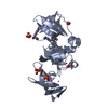
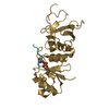
- PDB-5kco: SETDB1 in complex with an early stage, low affinity fragment cand... -
+ Open data

- Basic information
| Entry | Database: PDB / ID: 5kco | ||||||
|---|---|---|---|---|---|---|---|
| Title | SETDB1 in complex with an early stage, low affinity fragment candidate modelled at reduced occupancy | ||||||
 Components Components | Histone-lysine N-methyltransferase SETDB1 | ||||||
 Keywords Keywords | TRANSFERASE / Fragment Screening / DIAMOND I04-1 XCHEM / PANDDA / Structural Genomics / Structural Genomics Consortium / SGC | ||||||
| Function / homology |  Function and homology information Function and homology informationhistone H3K9 dimethyltransferase activity / [histone H3]-N6,N6-dimethyl-lysine9 N-methyltransferase / histone H3K9me2 methyltransferase activity / histone H3K9 trimethyltransferase activity / transposable element silencing by heterochromatin formation / histone H3K9 methyltransferase activity / histone H3K9 monomethyltransferase activity / heterochromatin organization / histone H3K14ac reader activity / histone H3K9me2/3 reader activity ...histone H3K9 dimethyltransferase activity / [histone H3]-N6,N6-dimethyl-lysine9 N-methyltransferase / histone H3K9me2 methyltransferase activity / histone H3K9 trimethyltransferase activity / transposable element silencing by heterochromatin formation / histone H3K9 methyltransferase activity / histone H3K9 monomethyltransferase activity / heterochromatin organization / histone H3K14ac reader activity / histone H3K9me2/3 reader activity / histone H3 methyltransferase activity / DNA methylation-dependent constitutive heterochromatin formation / Regulation of endogenous retroelements by the Human Silencing Hub (HUSH) complex / Regulation of endogenous retroelements by KRAB-ZFP proteins / promoter-specific chromatin binding / PKMTs methylate histone lysines / methylation / negative regulation of gene expression / chromatin binding / chromatin / DNA binding / zinc ion binding / nucleoplasm / nucleus / cytoplasm Similarity search - Function | ||||||
| Biological species |  Homo sapiens (human) Homo sapiens (human) | ||||||
| Method |  X-RAY DIFFRACTION / SYNCHROTRON / FOURIER SYNTHESIS / Resolution: 1.47 Å X-RAY DIFFRACTION / SYNCHROTRON / FOURIER SYNTHESIS / Resolution: 1.47 Å | ||||||
 Authors Authors | Tempel, W. / Harding, R.J. / Mader, P. / Dobrovetsky, E. / Walker, J.R. / Brown, P.J. / Schapira, M. / Collins, P. / Pearce, N. / Brandao-Neto, J. ...Tempel, W. / Harding, R.J. / Mader, P. / Dobrovetsky, E. / Walker, J.R. / Brown, P.J. / Schapira, M. / Collins, P. / Pearce, N. / Brandao-Neto, J. / Douangamath, A. / von Delft, F. / Bountra, C. / Arrowsmith, C.H. / Edwards, A.M. / Santhakumar, V. / Structural Genomics Consortium (SGC) | ||||||
 Citation Citation | Journal: To Be Published Title: SETDB1 in complex with an early stage, low affinity fragment candidate modelled at reduced occupancy Authors: Tempel, W. / Harding, R.J. / Mader, P. / Dobrovetsky, E. / Walker, J.R. / Brown, P.J. / Schapira, M. / Arrowsmith, C.H. / Edwards, A.M. / Santhakumar, S. / Structural Genomics Consortium (SGC) | ||||||
| History |
|
- Structure visualization
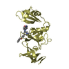
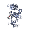
| Structure viewer | Molecule: MolmilJmol/JSmol |
|---|
- Downloads & links
-Download
| PDBx/mmCIF format | 5kco.cif.gz | 67.9 KB | Display | PDBx/mmCIF format |
|---|---|---|---|---|
| PDB format | pdb5kco.ent.gz | 47.8 KB | Display | PDB format |
| PDBx/mmJSON format | 5kco.json.gz | Tree view | PDBx/mmJSON format | |
| Others |  Other downloads Other downloads |
-Validation report
| Arichive directory | https://data.pdbj.org/pub/pdb/validation_reports/kc/5kcoftp://data.pdbj.org/pub/pdb/validation_reports/kc/5kco | HTTPS FTP |
|---|
-Related structure data
| Related structure data |  5kchC  3dlmS C: citing same article ( S: Starting model for refinement |
|---|---|
| Similar structure data |


-Links
 PDBj
PDBj
- Assembly
| Deposited unit | 
| ||||||||
|---|---|---|---|---|---|---|---|---|---|
| 1 |
| ||||||||
| Unit cell |
|
-Components
-Protein , 1 types, 1 molecules A
| #1: Protein | Mass: 26337.348 Da / Num. of mol.: 1 / Fragment: UNP residues 196-403 Source method: isolated from a genetically manipulated source Source: (gene. exp.) Homo sapiens (human) / Gene: SETDB1, KIAA0067, KMT1E / Plasmid: pET28-MHL / Production host:  References: UniProt: Q15047, histone-lysine N-methyltransferase |
|---|
-Non-polymers , 5 types, 196 molecules 
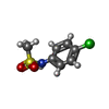


| #2: Chemical |  Mass: 96.063 Da / Num. of mol.: 2 / Source method: obtained synthetically / Formula: SO4 Mass: 96.063 Da / Num. of mol.: 2 / Source method: obtained synthetically / Formula: SO4#3: Chemical | ChemComp-6RO / ~{ |  Mass: 205.662 Da / Num. of mol.: 1 / Source method: obtained synthetically / Formula: C7H8ClNO2S Mass: 205.662 Da / Num. of mol.: 1 / Source method: obtained synthetically / Formula: C7H8ClNO2S#4: Chemical | ChemComp-UNX /  Mass: 96.063 Da / Num. of mol.: 47 / Source method: obtained synthetically Mass: 96.063 Da / Num. of mol.: 47 / Source method: obtained synthetically#5: Chemical |  Mass: 78.133 Da / Num. of mol.: 3 / Source method: obtained synthetically / Formula: C2H6OS / Comment: DMSO, precipitant*YM Mass: 78.133 Da / Num. of mol.: 3 / Source method: obtained synthetically / Formula: C2H6OS / Comment: DMSO, precipitant*YM#6: Water | ChemComp-HOH / | Mass: 18.015 Da / Num. of mol.: 143 / Source method: isolated from a natural source / Formula: H2O |
|---|
-Experimental details
-Experiment
| Experiment | Method: X-RAY DIFFRACTION / Number of used crystals: 1 |
|---|
- Sample preparation
| Crystal | Density Matthews: 2.38 Å3/Da / Density % sol: 48.33 % / Mosaicity: 0.06 ° |
|---|---|
| Crystal grow | Temperature: 291 K / Method: vapor diffusion, sitting drop / pH: 6.5 Details: 25% PEG-3350, 0.2M lithium sulfate, 0.1M bis-tris. Trypsin had been added to the protein stock solution. |
-Data collection
| Diffraction | Mean temperature: 100 K | ||||||||||||||||||||||||||||||
|---|---|---|---|---|---|---|---|---|---|---|---|---|---|---|---|---|---|---|---|---|---|---|---|---|---|---|---|---|---|---|---|
| Diffraction source | Source: SYNCHROTRON / Site: Diamond  / Beamline: I04-1 / Wavelength: 0.92819 Å / Beamline: I04-1 / Wavelength: 0.92819 Å | ||||||||||||||||||||||||||||||
| Detector | Type: DECTRIS PILATUS 6M-F / Detector: PIXEL / Date: Oct 11, 2015 | ||||||||||||||||||||||||||||||
| Radiation | Protocol: SINGLE WAVELENGTH / Monochromatic (M) / Laue (L): M / Scattering type: x-ray | ||||||||||||||||||||||||||||||
| Radiation wavelength | Wavelength: 0.92819 Å / Relative weight: 1 | ||||||||||||||||||||||||||||||
| Reflection | Resolution: 1.47→47.3 Å / Num. obs: 43355 / % possible obs: 99.7 % / Redundancy: 6.6 % / CC1/2: 0.998 / Rmerge(I) obs: 0.088 / Rpim(I) all: 0.037 / Rrim(I) all: 0.096 / Net I/σ(I): 11.8 / Num. measured all: 284319 | ||||||||||||||||||||||||||||||
| Reflection shell | Diffraction-ID: 1 / Rejects: _
|
- Processing
| Software |
| |||||||||||||||||||||||||||||||||||||||||||||||||||||||||||||||||||||||||||
|---|---|---|---|---|---|---|---|---|---|---|---|---|---|---|---|---|---|---|---|---|---|---|---|---|---|---|---|---|---|---|---|---|---|---|---|---|---|---|---|---|---|---|---|---|---|---|---|---|---|---|---|---|---|---|---|---|---|---|---|---|---|---|---|---|---|---|---|---|---|---|---|---|---|---|---|---|
| Refinement | Method to determine structure: FOURIER SYNTHESIS Starting model: 3DLM Resolution: 1.47→47.3 Å / Cor.coef. Fo:Fc: 0.963 / Cor.coef. Fo:Fc free: 0.956 / SU B: 1.422 / SU ML: 0.052 / Cross valid method: THROUGHOUT / σ(F): 0 / ESU R: 0.068 / ESU R Free: 0.071 / Stereochemistry target values: MAXIMUM LIKELIHOOD Details: Users of this crystal structure: verify our intepretion of the electron density. Amplitudes and unmerged intensities are included with this deposition. Diffraction images will be deposited ...Details: Users of this crystal structure: verify our intepretion of the electron density. Amplitudes and unmerged intensities are included with this deposition. Diffraction images will be deposited in a public repository. Geometry restraints for the fragment candidate were prepared with GRADE. Some occupancies were refined with PHENIX software. Ambiguous difference density suggests more than one main chain conformation for residues 235..239.
| |||||||||||||||||||||||||||||||||||||||||||||||||||||||||||||||||||||||||||
| Solvent computation | Ion probe radii: 0.8 Å / Shrinkage radii: 0.8 Å / VDW probe radii: 1.2 Å / Solvent model: MASK | |||||||||||||||||||||||||||||||||||||||||||||||||||||||||||||||||||||||||||
| Displacement parameters | Biso max: 66.74 Å2 / Biso mean: 20.634 Å2 / Biso min: 10.36 Å2
| |||||||||||||||||||||||||||||||||||||||||||||||||||||||||||||||||||||||||||
| Refinement step | Cycle: final / Resolution: 1.47→47.3 Å
| |||||||||||||||||||||||||||||||||||||||||||||||||||||||||||||||||||||||||||
| Refine LS restraints |
| |||||||||||||||||||||||||||||||||||||||||||||||||||||||||||||||||||||||||||
| LS refinement shell | Resolution: 1.47→1.508 Å / Total num. of bins used: 20
|
