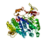
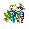
 Movie
Movie Controller


+ Open data

- Basic information
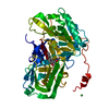
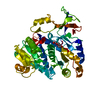
| Entry | Database: PDB / ID: 5gyz | ||||||
|---|---|---|---|---|---|---|---|
| Title | luciferase AMP/7-cy-L complex | ||||||
 Components Components | Luciferin 4-monooxygenase | ||||||
 Keywords Keywords | OXIDOREDUCTASE / Substrate / Luciferase | ||||||
| Function / homology |  Function and homology information Function and homology informationPhotinus-luciferin 4-monooxygenase (ATP-hydrolyzing) activity / firefly luciferase / CoA-ligase activity / bioluminescence / peroxisome / protein-folding chaperone binding / ATP binding / metal ion binding Similarity search - Function | ||||||
| Biological species |  Photinus pyralis (common eastern firefly) Photinus pyralis (common eastern firefly) | ||||||
| Method |  X-RAY DIFFRACTION / FREE ELECTRON LASER / MOLECULAR REPLACEMENT / Resolution: 2.4 Å X-RAY DIFFRACTION / FREE ELECTRON LASER / MOLECULAR REPLACEMENT / Resolution: 2.4 Å | ||||||
 Authors Authors | Su, J. / Wang, F. | ||||||
| Funding support |  China, 1items China, 1items
| ||||||
 Citation Citation | Journal: To Be Published Title: Structure of luciferase with AMP/7-cy-L at 2.4 Angstroms resolution Authors: Chao, T.Z. / Su, J. | ||||||
| History |
|
- Structure visualization
| Structure viewer | Molecule: MolmilJmol/JSmol |
|---|
- Downloads & links
-Download
| PDBx/mmCIF format | 5gyz.cif.gz | 106.8 KB | Display | PDBx/mmCIF format |
|---|---|---|---|---|
| PDB format | pdb5gyz.ent.gz | 78.7 KB | Display | PDB format |
| PDBx/mmJSON format | 5gyz.json.gz | Tree view | PDBx/mmJSON format | |
| Others |  Other downloads Other downloads |
-Validation report
| Arichive directory | https://data.pdbj.org/pub/pdb/validation_reports/gy/5gyzftp://data.pdbj.org/pub/pdb/validation_reports/gy/5gyz | HTTPS FTP |
|---|
-Related structure data
| Related structure data |  1ba3S S: Starting model for refinement |
|---|---|
| Similar structure data |


-Links
 PDBj
PDBj


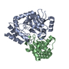
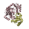
- Assembly
| Deposited unit | 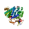
| ||||||||
|---|---|---|---|---|---|---|---|---|---|
| 1 |
| ||||||||
| Unit cell |
|
-Components
-Protein , 1 types, 1 molecules A
| #1: Protein | Mass: 48417.523 Da / Num. of mol.: 1 / Fragment: UNP residues 4-438 Source method: isolated from a genetically manipulated source Source: (gene. exp.) Photinus pyralis (common eastern firefly)Production host:  |
|---|
-Non-polymers , 5 types, 227 molecules 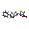


| #2: Chemical | ChemComp-7BV / ( Mass: 361.482 Da / Num. of mol.: 1 / Source method: obtained synthetically / Formula: C17H19N3O2S2 Mass: 361.482 Da / Num. of mol.: 1 / Source method: obtained synthetically / Formula: C17H19N3O2S2 | ||
|---|---|---|---|
| #3: Chemical | ChemComp-AMP /  Mass: 347.221 Da / Num. of mol.: 1 Mass: 347.221 Da / Num. of mol.: 1Source method: isolated from a genetically manipulated source Formula: C10H14N5O7P / Comment: AMP*YM | ||
| #4: Chemical | ChemComp-PEG /  Mass: 106.120 Da / Num. of mol.: 1 Mass: 106.120 Da / Num. of mol.: 1Source method: isolated from a genetically manipulated source Formula: C4H10O3 | ||
| #5: Chemical |  Mass: 92.094 Da / Num. of mol.: 3 Mass: 92.094 Da / Num. of mol.: 3Source method: isolated from a genetically manipulated source Formula: C3H8O3 #6: Water | ChemComp-HOH / | Mass: 18.015 Da / Num. of mol.: 221 / Source method: isolated from a natural source / Formula: H2O |
-Experimental details
-Experiment
| Experiment | Method: X-RAY DIFFRACTION / Number of used crystals: 1 |
|---|
- Sample preparation
| Crystal | Density Matthews: 2.68 Å3/Da / Density % sol: 54.03 % |
|---|---|
| Crystal grow | Temperature: 293 K / Method: evaporation / Details: Sodium malonate, PEG3350 |
-Data collection
| Diffraction | Mean temperature: 100 K |
|---|---|
| Diffraction source | Source: FREE ELECTRON LASER / Site: SACLA  / Beamline: BL3 / Wavelength: 0.9791 Å / Beamline: BL3 / Wavelength: 0.9791 Å |
| Detector | Type: MARMOSAIC 225 mm CCD / Detector: CCD / Date: Apr 20, 2016 |
| Radiation | Protocol: SINGLE WAVELENGTH / Monochromatic (M) / Laue (L): M / Scattering type: x-ray |
| Radiation wavelength | Wavelength: 0.9791 Å / Relative weight: 1 |
| Reflection | Resolution: 2.4→50 Å / Num. obs: 19770 / % possible obs: 98.9 % / Redundancy: 7.4 % / Net I/σ(I): 47.1 |
- Processing
| Software |
| ||||||||||||||||||||||||||||||||||||||||||||||||||||||||
|---|---|---|---|---|---|---|---|---|---|---|---|---|---|---|---|---|---|---|---|---|---|---|---|---|---|---|---|---|---|---|---|---|---|---|---|---|---|---|---|---|---|---|---|---|---|---|---|---|---|---|---|---|---|---|---|---|---|
| Refinement | Method to determine structure: MOLECULAR REPLACEMENT Starting model: 1BA3 Resolution: 2.4→40.2 Å / SU ML: 0.28 / Cross valid method: FREE R-VALUE / σ(F): 1.34 / Phase error: 24.82
| ||||||||||||||||||||||||||||||||||||||||||||||||||||||||
| Solvent computation | Shrinkage radii: 0.9 Å / VDW probe radii: 1.11 Å | ||||||||||||||||||||||||||||||||||||||||||||||||||||||||
| Refinement step | Cycle: LAST / Resolution: 2.4→40.2 Å
| ||||||||||||||||||||||||||||||||||||||||||||||||||||||||
| Refine LS restraints |
| ||||||||||||||||||||||||||||||||||||||||||||||||||||||||
| LS refinement shell |
|
