spontaneous exocytosis of neurotransmitter / PKA activation in glucagon signalling / CREB1 phosphorylation through the activation of Adenylate Cyclase / negative regulation of meiotic cell cycle / HDL assembly / DARPP-32 events / Rap1 signalling / PKA activation / Vasopressin regulates renal water homeostasis via Aquaporins / Regulation of insulin secretion ...spontaneous exocytosis of neurotransmitter / PKA activation in glucagon signalling / CREB1 phosphorylation through the activation of Adenylate Cyclase / negative regulation of meiotic cell cycle / HDL assembly / DARPP-32 events / Rap1 signalling / PKA activation / Vasopressin regulates renal water homeostasis via Aquaporins / Regulation of insulin secretion / GPER1 signaling / Hedgehog 'off' state / Glucagon-like Peptide-1 (GLP1) regulates insulin secretion / regulation of membrane repolarization / exocytic vesicle / Loss of Nlp from mitotic centrosomes / Recruitment of mitotic centrosome proteins and complexes / Loss of proteins required for interphase microtubule organization from the centrosome / Recruitment of NuMA to mitotic centrosomes / Anchoring of the basal body to the plasma membrane / MAPK6/MAPK4 signaling / GLI3 is processed to GLI3R by the proteasome / AURKA Activation by TPX2 / Factors involved in megakaryocyte development and platelet production / cAMP-dependent protein kinase regulator activity / Regulation of PLK1 Activity at G2/M Transition / Interleukin-3, Interleukin-5 and GM-CSF signaling / CD209 (DC-SIGN) signaling / Mitochondrial protein degradation / positive regulation of potassium ion transmembrane transport / RET signaling / nucleotide-activated protein kinase complex / regulation of protein kinase A signaling / Ion homeostasis / VEGFA-VEGFR2 Pathway / regulation of cellular respiration / regulation of protein processing / protein localization to lipid droplet / regulation of bicellular tight junction assembly / cellular response to parathyroid hormone stimulus / cAMP-dependent protein kinase inhibitor activity / cAMP-dependent protein kinase / cellular response to cold / protein kinase A binding / regulation of osteoblast differentiation / sperm capacitation / cAMP-dependent protein kinase activity / ciliary base / negative regulation of glycolytic process through fructose-6-phosphate / cAMP-dependent protein kinase complex / AMP-activated protein kinase activity / postsynaptic modulation of chemical synaptic transmission / cellular response to glucagon stimulus / AMP binding / protein kinase A regulatory subunit binding / plasma membrane raft / protein kinase A catalytic subunit binding / axoneme / mesoderm formation / lateral plasma membrane / small molecule binding / sperm flagellum / negative regulation of smoothened signaling pathway / regulation of proteasomal protein catabolic process / beta-2 adrenergic receptor binding / cAMP binding / cellular response to cAMP / regulation of synaptic transmission, glutamatergic / positive regulation of gluconeogenesis / sperm midpiece / negative regulation of TORC1 signaling / T-tubule / protein kinase A signaling / protein serine/threonine/tyrosine kinase activity / protein export from nucleus / hippocampal mossy fiber to CA3 synapse / positive regulation of protein export from nucleus / acrosomal vesicle / sarcoplasmic reticulum / neural tube closure / cellular response to glucose stimulus / regulation of protein phosphorylation / modulation of chemical synaptic transmission / neuromuscular junction / adenylate cyclase-activating G protein-coupled receptor signaling pathway / positive regulation of insulin secretion / mRNA processing / small GTPase binding / presynapse / kinase activity / manganese ion binding / cellular response to heat / postsynapse / peptidyl-serine phosphorylation / dendritic spine / regulation of cell cycle / protein kinase activity / nuclear speck / phosphorylation / apical plasma membrane 類似検索 - 分子機能
A-kinase anchor protein 7, RI-RII subunit-binding domain / PKA-RI-RII subunit binding domain of A-kinase anchor protein / Protein kinase A anchor protein, nuclear localisation signal domain / AKAP7 2'5' RNA ligase-like domain / cAMP-dependent protein kinase regulatory subunit / cAMP-dependent protein kinase regulatory subunit, dimerization-anchoring domain / Regulatory subunit of type II PKA R-subunit / RIIalpha, Regulatory subunit portion of type II PKA R-subunit / Cyclic phosphodiesterase / Cyclic nucleotide-binding domain signature 2. ...A-kinase anchor protein 7, RI-RII subunit-binding domain / PKA-RI-RII subunit binding domain of A-kinase anchor protein / Protein kinase A anchor protein, nuclear localisation signal domain / AKAP7 2'5' RNA ligase-like domain / cAMP-dependent protein kinase regulatory subunit / cAMP-dependent protein kinase regulatory subunit, dimerization-anchoring domain / Regulatory subunit of type II PKA R-subunit / RIIalpha, Regulatory subunit portion of type II PKA R-subunit / Cyclic phosphodiesterase / Cyclic nucleotide-binding domain signature 2. / Cyclic nucleotide-binding domain signature 1. / cAMP-dependent protein kinase catalytic subunit / Cyclic nucleotide-binding, conserved site / Cyclic nucleotide-monophosphate binding domain / Cyclic nucleotide-binding domain / cAMP/cGMP binding motif profile. / Cyclic nucleotide-binding domain / Cyclic nucleotide-binding domain superfamily / Extension to Ser/Thr-type protein kinases / AGC-kinase, C-terminal / AGC-kinase C-terminal domain profile. / RmlC-like jelly roll fold / Serine/threonine-protein kinase, active site / Serine/Threonine protein kinases active-site signature. / Protein kinase domain / Serine/Threonine protein kinases, catalytic domain / Protein kinase, ATP binding site / Protein kinases ATP-binding region signature. / Protein kinase domain profile. / Protein kinase domain / Protein kinase-like domain superfamily 類似検索 - ドメイン・相同性
cAMP-dependent protein kinase catalytic subunit alpha / cAMP-dependent protein kinase type II-alpha regulatory subunit / A-kinase anchor protein 7 isoforms delta and gamma / cAMP-dependent protein kinase type II-alpha regulatory subunit 類似検索 - 構成要素
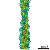
ジャーナル: Elife / 年: 2013 タイトル: Intrinsic disorder within an AKAP-protein kinase A complex guides local substrate phosphorylation. 著者: F Donelson Smith / Steve L Reichow / Jessica L Esseltine / Dan Shi / Lorene K Langeberg / John D Scott / Tamir Gonen / 要旨: Anchoring proteins sequester kinases with their substrates to locally disseminate intracellular signals and avert indiscriminate transmission of these responses throughout the cell. Mechanistic ...Anchoring proteins sequester kinases with their substrates to locally disseminate intracellular signals and avert indiscriminate transmission of these responses throughout the cell. Mechanistic understanding of this process is hampered by limited structural information on these macromolecular complexes. A-kinase anchoring proteins (AKAPs) spatially constrain phosphorylation by cAMP-dependent protein kinases (PKA). Electron microscopy and three-dimensional reconstructions of type-II PKA-AKAP18γ complexes reveal hetero-pentameric assemblies that adopt a range of flexible tripartite configurations. Intrinsically disordered regions within each PKA regulatory subunit impart the molecular plasticity that affords an ∼16 nanometer radius of motion to the associated catalytic subunits. Manipulating flexibility within the PKA holoenzyme augmented basal and cAMP responsive phosphorylation of AKAP-associated substrates. Cell-based analyses suggest that the catalytic subunit remains within type-II PKA-AKAP18γ complexes upon cAMP elevation. We propose that the dynamic movement of kinase sub-structures, in concert with the static AKAP-regulatory subunit interface, generates a solid-state signaling microenvironment for substrate phosphorylation. DOI: http://dx.doi.org/10.7554/eLife.01319.001.
タンパク質・ペプチド: A-Protein Kinase Regulatory Subunit II alpha
-
超分子 #1000: AKAP18-PKA Complex in Linear Conformation
超分子
名称: AKAP18-PKA Complex in Linear Conformation / タイプ: sample / ID: 1000 集合状態: Hetero-pentamer composed of one AKAP18 bound to a dimer of the PKA Regulatory Subunit II and two copies of the PKA Catalytic Subunit Number unique components: 3
 ムービー
ムービー コントローラー


 データを開く
 基本情報
基本情報 マップデータ
 試料
試料 キーワード
キーワード 機能・相同性情報
 Homo sapiens (ヒト) /
 データ登録者
 引用
引用
 構造の表示
構造の表示
 ダウンロードとリンク
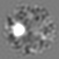
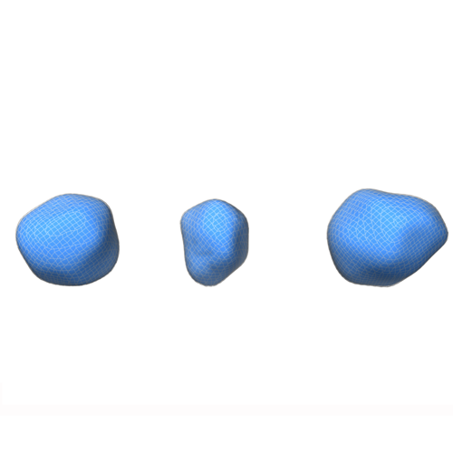 emd_5756.png
 http://ftp.pdbj.org/pub/emdb/structures/EMD-5756
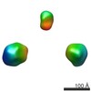


 Z (Sec.)
 Y (Row.)
 X (Col.)


 試料の構成要素
 Spodoptera frugiperda (ツマジロクサヨトウ)
 解析
解析 電子顕微鏡法
 Chimera


