 Movie
Movie Controller

[English] 日本語
 Yorodumi
- PDB-3j4r: Pseudo-atomic model of the AKAP18-PKA Complex in a linear conform... -
+ Open data

- Basic information
| Entry | Database: PDB / ID: 3j4r | ||||||
|---|---|---|---|---|---|---|---|
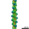
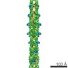
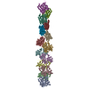
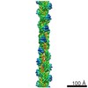
| Title | Pseudo-atomic model of the AKAP18-PKA Complex in a linear conformation derived from electron microscopy | ||||||
 Components Components |
| ||||||
 Keywords Keywords | TRANSFERASE / A-kinase anchoring protein / cAMP-Dependent Kinase / RII / PKA regulatory subunit II / phosphorylation / anchoring / intrinsic disorder | ||||||
| Function / homology |  Function and homology information Function and homology informationspontaneous exocytosis of neurotransmitter / PKA activation in glucagon signalling / CREB1 phosphorylation through the activation of Adenylate Cyclase / negative regulation of meiotic cell cycle / HDL assembly / DARPP-32 events / Rap1 signalling / regulation of protein kinase activity / PKA activation / Regulation of insulin secretion ...spontaneous exocytosis of neurotransmitter / PKA activation in glucagon signalling / CREB1 phosphorylation through the activation of Adenylate Cyclase / negative regulation of meiotic cell cycle / HDL assembly / DARPP-32 events / Rap1 signalling / regulation of protein kinase activity / PKA activation / Regulation of insulin secretion / Vasopressin regulates renal water homeostasis via Aquaporins / GPER1 signaling / Hedgehog 'off' state / Glucagon-like Peptide-1 (GLP1) regulates insulin secretion / Loss of Nlp from mitotic centrosomes / Recruitment of mitotic centrosome proteins and complexes / Loss of proteins required for interphase microtubule organization from the centrosome / positive regulation of triglyceride catabolic process / exocytic vesicle / Anchoring of the basal body to the plasma membrane / Recruitment of NuMA to mitotic centrosomes / AURKA Activation by TPX2 / cAMP-dependent protein kinase regulator activity / Factors involved in megakaryocyte development and platelet production / MAPK6/MAPK4 signaling / regulation of cellular respiration / Interleukin-3, Interleukin-5 and GM-CSF signaling / CD209 (DC-SIGN) signaling / regulation of membrane repolarization / RET signaling / GLI3 is processed to GLI3R by the proteasome / High laminar flow shear stress activates signaling by PIEZO1 and PECAM1:CDH5:KDR in endothelial cells / Regulation of PLK1 Activity at G2/M Transition / Mitochondrial protein degradation / VEGFA-VEGFR2 Pathway / nucleotide-activated protein kinase complex / Ion homeostasis / positive regulation of potassium ion transmembrane transport / regulation of protein processing / cAMP-dependent protein kinase inhibitor activity / cellular response to parathyroid hormone stimulus / protein localization to lipid droplet / potassium channel inhibitor activity / histone H1-4S35 kinase activity / cAMP-dependent protein kinase / beta-2 adrenergic receptor binding / negative regulation of glycolytic process / cAMP-dependent protein kinase activity / regulation of bicellular tight junction assembly / cAMP-dependent protein kinase complex / mesoderm formation / cellular response to cold / AMP binding / interleukin-2-mediated signaling pathway / sperm capacitation / regulation of osteoblast differentiation / peptidyl-threonine phosphorylation / negative regulation of glycolytic process through fructose-6-phosphate / protein kinase A regulatory subunit binding / ciliary base / protein kinase A catalytic subunit binding / small molecule binding / TORC1 signaling / protein kinase A binding / intracellular potassium ion homeostasis / action potential / lateral plasma membrane / plasma membrane raft / cAMP/PKA signal transduction / axoneme / neural tube closure / sperm flagellum / postsynaptic modulation of chemical synaptic transmission / cAMP binding / negative regulation of cAMP/PKA signal transduction / regulation of synaptic transmission, glutamatergic / negative regulation of TORC1 signaling / positive regulation of gluconeogenesis / protein serine/threonine/tyrosine kinase activity / negative regulation of protein localization to chromatin / cellular response to glucagon stimulus / cellular response to nutrient levels / lipid droplet / T-tubule / positive regulation of phagocytosis / positive regulation of protein export from nucleus / acrosomal vesicle / cellular response to cAMP / regulation of proteasomal protein catabolic process / hippocampal mossy fiber to CA3 synapse / negative regulation of smoothened signaling pathway / regulation of microtubule cytoskeleton organization / peptidyl-serine phosphorylation / sarcoplasmic reticulum / cellular response to glucose stimulus / neuromuscular junction / positive regulation of cholesterol biosynthetic process / modulation of chemical synaptic transmission / small GTPase binding / mRNA processing Similarity search - Function | ||||||
| Biological species |  Homo sapiens (human) Homo sapiens (human) | ||||||
| Method | ELECTRON MICROSCOPY / single particle reconstruction / negative staining / Resolution: 35 Å | ||||||
 Authors Authors | Reichow, S.L. / Gonen, T. | ||||||
 Citation Citation | Journal: Elife / Year: 2013 Title: Intrinsic disorder within an AKAP-protein kinase A complex guides local substrate phosphorylation. Authors: F Donelson Smith / Steve L Reichow / Jessica L Esseltine / Dan Shi / Lorene K Langeberg / John D Scott / Tamir Gonen /  Abstract: Anchoring proteins sequester kinases with their substrates to locally disseminate intracellular signals and avert indiscriminate transmission of these responses throughout the cell. Mechanistic ...Anchoring proteins sequester kinases with their substrates to locally disseminate intracellular signals and avert indiscriminate transmission of these responses throughout the cell. Mechanistic understanding of this process is hampered by limited structural information on these macromolecular complexes. A-kinase anchoring proteins (AKAPs) spatially constrain phosphorylation by cAMP-dependent protein kinases (PKA). Electron microscopy and three-dimensional reconstructions of type-II PKA-AKAP18γ complexes reveal hetero-pentameric assemblies that adopt a range of flexible tripartite configurations. Intrinsically disordered regions within each PKA regulatory subunit impart the molecular plasticity that affords an ∼16 nanometer radius of motion to the associated catalytic subunits. Manipulating flexibility within the PKA holoenzyme augmented basal and cAMP responsive phosphorylation of AKAP-associated substrates. Cell-based analyses suggest that the catalytic subunit remains within type-II PKA-AKAP18γ complexes upon cAMP elevation. We propose that the dynamic movement of kinase sub-structures, in concert with the static AKAP-regulatory subunit interface, generates a solid-state signaling microenvironment for substrate phosphorylation. DOI: http://dx.doi.org/10.7554/eLife.01319.001. | ||||||
| History |
|
- Structure visualization
| Movie |
Movie viewer |
|---|---|
| Structure viewer | Molecule: MolmilJmol/JSmol |
 UCSF Chimera
- Downloads & links
-Download
| PDBx/mmCIF format | 3j4r.cif.gz | 323.9 KB | Display | PDBx/mmCIF format |
|---|---|---|---|---|
| PDB format | pdb3j4r.ent.gz | 259 KB | Display | PDB format |
| PDBx/mmJSON format | 3j4r.json.gz | Tree view | PDBx/mmJSON format | |
| Others |  Other downloads Other downloads |
-Validation report
| Arichive directory | https://data.pdbj.org/pub/pdb/validation_reports/j4/3j4rftp://data.pdbj.org/pub/pdb/validation_reports/j4/3j4r | HTTPS FTP |
|---|
-Related structure data
| Related structure data |  5756MC 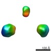 5755C  3j4qC M: map data used to model this data C: citing same article ( |
|---|---|
| Similar structure data |
-Links
 PDBj
PDBj


- Assembly
| Deposited unit | 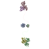
|
|---|---|
| 1 |
|
-Components
| #1: Protein | Mass: 39478.773 Da / Num. of mol.: 1 / Fragment: SEE REMARK 999 Source method: isolated from a genetically manipulated source Source: (gene. exp.) Homo sapiens (human) / Production host:   Spodoptera frugiperda (fall armyworm) / Strain (production host): Sf-9 / References: UniProt: Q6JP77*PLUS Spodoptera frugiperda (fall armyworm) / Strain (production host): Sf-9 / References: UniProt: Q6JP77*PLUS | ||||
|---|---|---|---|---|---|
| #2: Protein | Mass: 45644.266 Da / Num. of mol.: 2 Source method: isolated from a genetically manipulated source Source: (gene. exp.)  #3: Protein | Mass: 40628.555 Da / Num. of mol.: 2 Source method: isolated from a genetically manipulated source Source: (gene. exp.) Sequence details | AKAP18 IN THIS ENTRY IS FROM HOMO SAPIENS, BUT THE MODELED SEQUENCE IS FROM RATTUS NORVEGICUS | |
-Experimental details
-Experiment
| Experiment | Method: ELECTRON MICROSCOPY |
|---|---|
| EM experiment | Aggregation state: PARTICLE / 3D reconstruction method: single particle reconstruction |
- Sample preparation
| Component |
| |||||||||||||||||||||||||
|---|---|---|---|---|---|---|---|---|---|---|---|---|---|---|---|---|---|---|---|---|---|---|---|---|---|---|
| Molecular weight |
| |||||||||||||||||||||||||
| Buffer solution | Name: 25 mM HEPES, pH 7.4, 200 mM NaCl, 0.5 mM EDTA, 1 mM DTT pH: 7.4 Details: 25 mM HEPES, pH 7.4, 200 mM NaCl, 0.5 mM EDTA, 1 mM DTT | |||||||||||||||||||||||||
| Specimen | Conc.: 0.005 mg/ml / Embedding applied: NO / Shadowing applied: NO / Staining applied: YES / Vitrification applied: NO / Details: Stain 0.75% (w/v) uranyl formate | |||||||||||||||||||||||||
| EM staining | Type: NEGATIVE / Material: Uranyl Formate | |||||||||||||||||||||||||
| Specimen support | Details: 200 mesh copper grid with thin carbon support |
- Electron microscopy imaging
| Microscopy | Model: FEI TECNAI 12 / Date: May 1, 2012 |
|---|---|
| Electron gun | Electron source: LAB6 / Accelerating voltage: 120 kV / Illumination mode: FLOOD BEAM |
| Electron lens | Mode: BRIGHT FIELD / Nominal magnification: 52000 X / Nominal defocus max: 2500 nm / Nominal defocus min: 1000 nm / Cs: 6.3 mm Astigmatism: Objective lens astigmatism was corrected at 100,000 times magnification Camera length: 0 mm |
| Specimen holder | Specimen holder model: OTHER / Specimen holder type: Single-Tilt / Temperature: 298 K / Tilt angle max: 50 ° / Tilt angle min: 0 ° |
| Image recording | Electron dose: 15 e/Å2 / Film or detector model: GENERIC GATAN |
| Image scans | Num. digital images: 1335 |
| Radiation | Protocol: SINGLE WAVELENGTH / Monochromatic (M) / Laue (L): M / Scattering type: x-ray |
| Radiation wavelength | Relative weight: 1 |
- Processing
| EM software |
| ||||||||||||||||||||||||||||||||||||
|---|---|---|---|---|---|---|---|---|---|---|---|---|---|---|---|---|---|---|---|---|---|---|---|---|---|---|---|---|---|---|---|---|---|---|---|---|---|
| CTF correction | Details: Each Micrograph | ||||||||||||||||||||||||||||||||||||
| Symmetry | Point symmetry: C1 (asymmetric) | ||||||||||||||||||||||||||||||||||||
| 3D reconstruction | Method: Angular Reconstitution and Refinement / Resolution: 35 Å / Resolution method: FSC 0.5 CUT-OFF / Num. of particles: 1000 / Nominal pixel size: 4.2 Å / Actual pixel size: 4.2 Å Details: (Single particle details: Images were processed in IMAGIC and ISAC. 3D reconstruction was done in IMAGIC and FREALIGN.) (Single particle--Applied symmetry: C1) Symmetry type: POINT | ||||||||||||||||||||||||||||||||||||
| Atomic model building |
| ||||||||||||||||||||||||||||||||||||
| Atomic model building | Source name: PDB / Type: experimental model
| ||||||||||||||||||||||||||||||||||||
| Refinement step | Cycle: LAST
|


