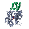
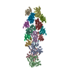
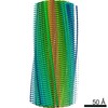
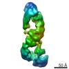
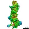
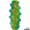
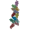
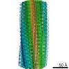
- EMDB-5170: Binding of alpha-actinin CH1 to F-actin -
+
データを開く
IDまたはキーワード:
読み込み中...
-
基本情報
登録情報
データベース: EMDB / ID: EMD-5170
タイトル
Binding of alpha-actinin CH1 to F-actin
マップデータ
reconstructed volume of F-actin decorated with alpha-actinin ABD (CH1 and CH2)
試料
試料: F-actin decorated with alpha-actinin ABD (containing CH1 and CH2)
タンパク質・ペプチド: F-actin
キーワード
helical filaments / calponin homology domains
機能・相同性
機能・相同性情報
positive regulation of glucose catabolic process to lactate via pyruvate / negative regulation of relaxation of muscle / skeletal muscle atrophy / regulation of the force of skeletal muscle contraction / positive regulation of skeletal muscle fiber development / positive regulation of skeletal muscle tissue growth / response to denervation involved in regulation of muscle adaptation / positive regulation of fast-twitch skeletal muscle fiber contraction / positive regulation of norepinephrine uptake / Regulation of CDH1 Function ...positive regulation of glucose catabolic process to lactate via pyruvate / negative regulation of relaxation of muscle / skeletal muscle atrophy / regulation of the force of skeletal muscle contraction / positive regulation of skeletal muscle fiber development / positive regulation of skeletal muscle tissue growth / response to denervation involved in regulation of muscle adaptation / positive regulation of fast-twitch skeletal muscle fiber contraction / positive regulation of norepinephrine uptake / Regulation of CDH1 Function / Formation of the polybromo-BAF (pBAF) complex / Formation of the canonical BAF (cBAF) complex / Formation of the non-canonical BAF (ncBAF) complex / Formation of the embryonic stem cell BAF (esBAF) complex / Formation of neuronal progenitor and neuronal BAF (npBAF and nBAF) / positive regulation of bone mineralization involved in bone maturation / bBAF complex / cellular response to cytochalasin B / npBAF complex / nBAF complex / brahma complex / regulation of transepithelial transport / morphogenesis of a polarized epithelium / Formation of annular gap junctions / Formation of the dystrophin-glycoprotein complex (DGC) / focal adhesion assembly / structural constituent of postsynaptic actin cytoskeleton / GBAF complex / transition between fast and slow fiber / Gap junction degradation / Folding of actin by CCT/TriC / regulation of G0 to G1 transition / Cell-extracellular matrix interactions / protein localization to adherens junction / muscle cell development / dense body / Tat protein binding / negative regulation of oxidative phosphorylation / postsynaptic actin cytoskeleton / Prefoldin mediated transfer of substrate to CCT/TriC / RSC-type complex / Striated Muscle Contraction / bone morphogenesis / regulation of nucleotide-excision repair / regulation of double-strand break repair / negative regulation of glycolytic process / Nephrin family interactions / Adherens junctions interactions / RHOF GTPase cycle / adherens junction assembly / negative regulation of cold-induced thermogenesis / apical protein localization / Sensory processing of sound by inner hair cells of the cochlea / Sensory processing of sound by outer hair cells of the cochlea / negative regulation of calcineurin-NFAT signaling cascade / Interaction between L1 and Ankyrins / tight junction / SWI/SNF complex / regulation of mitotic metaphase/anaphase transition / structural constituent of muscle / regulation of aerobic respiration / positive regulation of T cell differentiation / apical junction complex / cortical actin cytoskeleton / positive regulation of double-strand break repair / maintenance of blood-brain barrier / regulation of norepinephrine uptake / nitric-oxide synthase binding / transporter regulator activity / cortical cytoskeleton / positive regulation of stem cell population maintenance / establishment or maintenance of cell polarity / pseudopodium / NuA4 histone acetyltransferase complex / Recycling pathway of L1 / Regulation of MITF-M-dependent genes involved in pigmentation / brush border / regulation of G1/S transition of mitotic cell cycle / EPH-ephrin mediated repulsion of cells / negative regulation of cell differentiation / kinesin binding / RHO GTPases Activate WASPs and WAVEs / regulation of synaptic vesicle endocytosis / positive regulation of myoblast differentiation / RHO GTPases activate IQGAPs / regulation of protein localization to plasma membrane / positive regulation of double-strand break repair via homologous recombination / EPHB-mediated forward signaling / cytoskeleton organization / substantia nigra development / axonogenesis / calyx of Held / nitric-oxide synthase regulator activity / cell projection / FCGR3A-mediated phagocytosis / actin filament / adherens junction / Translocation of SLC2A4 (GLUT4) to the plasma membrane / positive regulation of cell differentiation / Regulation of endogenous retroelements by Piwi-interacting RNAs (piRNAs) 類似検索 - 分子機能
ジャーナル: Nat Struct Mol Biol / 年: 2010 タイトル: Opening of tandem calponin homology domains regulates their affinity for F-actin. 著者: Vitold E Galkin / Albina Orlova / Anita Salmazo / Kristina Djinovic-Carugo / Edward H Egelman / 要旨: Many actin-binding proteins contain calponin homology (CH) domains, but the manner in which these domains interact with F-actin has been controversial. Crystal structures have shown the tandem CH ...Many actin-binding proteins contain calponin homology (CH) domains, but the manner in which these domains interact with F-actin has been controversial. Crystal structures have shown the tandem CH domains of alpha-actinin to be in a compact, closed conformation, but the interpretations of complexes of such tandem CH domains with F-actin have been ambiguous. We show that the tandem CH domains of alpha-actinin bind F-actin in an open conformation, explaining mutations that cause human diseases and suggesting that the opening of these domains may be one of the main regulatory mechanisms for proteins with tandem CH domains.
全体 : F-actin decorated with alpha-actinin ABD (containing CH1 and CH2)
全体
名称: F-actin decorated with alpha-actinin ABD (containing CH1 and CH2)
要素
試料: F-actin decorated with alpha-actinin ABD (containing CH1 and CH2)
タンパク質・ペプチド: F-actin
-
超分子 #1000: F-actin decorated with alpha-actinin ABD (containing CH1 and CH2)
超分子
名称: F-actin decorated with alpha-actinin ABD (containing CH1 and CH2) タイプ: sample / ID: 1000 / 詳細: none / 集合状態: one to one binding / Number unique components: 2
 ムービー
ムービー コントローラー


 データを開く
 基本情報
基本情報 マップデータ
 試料
試料 キーワード
キーワード 機能・相同性情報
 Homo sapiens (ヒト)
 データ登録者
 引用
引用
 構造の表示
構造の表示
 ダウンロードとリンク
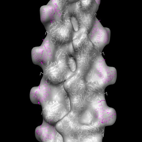 emd_5170_1.jpg
 http://ftp.pdbj.org/pub/emdb/structures/EMD-5170


 Z (Sec.)
 Y (Row.)
 X (Col.)


 試料の構成要素
 解析
解析 電子顕微鏡法
 FIELD EMISSION GUN