 Movie
Movie Controller

[English] 日本語
 Yorodumi
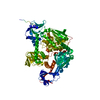
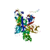
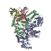
- PDB-4cak: Three-dimensional reconstruction of intact human integrin alphaII... -
+ Open data

- Basic information
| Entry | Database: PDB / ID: 4cak | |||||||||||||||
|---|---|---|---|---|---|---|---|---|---|---|---|---|---|---|---|---|
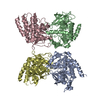
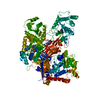
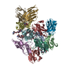
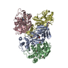
| Title | Three-dimensional reconstruction of intact human integrin alphaIIbbeta3 in a phospholipid bilayer nanodisc | |||||||||||||||
 Components Components |
| |||||||||||||||
 Keywords Keywords | CELL ADHESION / INTEGRIN / SINGLE PARTICLE RECONSTRUCTION | |||||||||||||||
| Function / homology |  Function and homology information Function and homology informationregulation of serotonin uptake / positive regulation of adenylate cyclase-inhibiting opioid receptor signaling pathway / tube development / alpha9-beta1 integrin-ADAM8 complex / regulation of trophoblast cell migration / integrin alphaIIb-beta3 complex / regulation of postsynaptic neurotransmitter receptor diffusion trapping / maintenance of postsynaptic specialization structure / alphav-beta3 integrin-vitronectin complex / regulation of extracellular matrix organization ...regulation of serotonin uptake / positive regulation of adenylate cyclase-inhibiting opioid receptor signaling pathway / tube development / alpha9-beta1 integrin-ADAM8 complex / regulation of trophoblast cell migration / integrin alphaIIb-beta3 complex / regulation of postsynaptic neurotransmitter receptor diffusion trapping / maintenance of postsynaptic specialization structure / alphav-beta3 integrin-vitronectin complex / regulation of extracellular matrix organization / platelet alpha granule membrane / positive regulation of glomerular mesangial cell proliferation / integrin alphav-beta3 complex / negative regulation of lipoprotein metabolic process / alphav-beta3 integrin-PKCalpha complex / fibrinogen binding / alphav-beta3 integrin-HMGB1 complex / negative regulation of lipid transport / vascular endothelial growth factor receptor 2 binding / positive regulation of vascular endothelial growth factor signaling pathway / Elastic fibre formation / cell-substrate junction assembly / alphav-beta3 integrin-IGF-1-IGF1R complex / positive regulation of bone resorption / platelet-derived growth factor receptor binding / angiogenesis involved in wound healing / mesodermal cell differentiation / filopodium membrane / regulation of release of sequestered calcium ion into cytosol / glycinergic synapse / extracellular matrix binding / positive regulation of vascular endothelial growth factor receptor signaling pathway / positive regulation of cell adhesion mediated by integrin / apolipoprotein A-I-mediated signaling pathway / regulation of bone resorption / negative regulation of low-density lipoprotein particle clearance / wound healing, spreading of epidermal cells / positive regulation of leukocyte migration / apoptotic cell clearance / positive regulation of fibroblast migration / integrin complex / cell adhesion mediated by integrin / smooth muscle cell migration / Molecules associated with elastic fibres / heterotypic cell-cell adhesion / positive regulation of smooth muscle cell migration / negative chemotaxis / Mechanical load activates signaling by PIEZO1 and integrins in osteocytes / positive regulation of cell-matrix adhesion / Syndecan interactions / p130Cas linkage to MAPK signaling for integrins / cellular response to insulin-like growth factor stimulus / regulation of postsynaptic neurotransmitter receptor internalization / positive regulation of osteoblast proliferation / protein disulfide isomerase activity / microvillus membrane / platelet-derived growth factor receptor signaling pathway / cell-substrate adhesion / PECAM1 interactions / GRB2:SOS provides linkage to MAPK signaling for Integrins / TGF-beta receptor signaling activates SMADs / lamellipodium membrane / fibronectin binding / negative regulation of macrophage derived foam cell differentiation / negative regulation of lipid storage / blood coagulation, fibrin clot formation / ECM proteoglycans / Integrin cell surface interactions / negative regulation of endothelial cell apoptotic process / positive regulation of T cell migration / coreceptor activity / cellular response to platelet-derived growth factor stimulus / Integrin signaling / positive regulation of endothelial cell proliferation / positive regulation of substrate adhesion-dependent cell spreading / substrate adhesion-dependent cell spreading / embryo implantation / cell adhesion molecule binding / positive regulation of endothelial cell migration / positive regulation of smooth muscle cell proliferation / Turbulent (oscillatory, disturbed) flow shear stress activates signaling by PIEZO1 and integrins in endothelial cells / cell-matrix adhesion / protein kinase C binding / response to activity / Signal transduction by L1 / integrin-mediated signaling pathway / regulation of actin cytoskeleton organization / wound healing / cellular response to mechanical stimulus / Signaling by high-kinase activity BRAF mutants / cell-cell adhesion / RUNX1 regulates genes involved in megakaryocyte differentiation and platelet function / platelet activation / MAP2K and MAPK activation / VEGFA-VEGFR2 Pathway / platelet aggregation / integrin binding / cellular response to xenobiotic stimulus / positive regulation of fibroblast proliferation / ruffle membrane Similarity search - Function | |||||||||||||||
| Biological species |  Homo sapiens (human) Homo sapiens (human) | |||||||||||||||
| Method | ELECTRON MICROSCOPY / single particle reconstruction / negative staining / Resolution: 20.5 Å | |||||||||||||||
 Authors Authors | Choi, W.S. / Rice, W.J. / Stokes, D.L. / Coller, B.S. | |||||||||||||||
 Citation Citation | Journal: Blood / Year: 2013 Title: Three-dimensional reconstruction of intact human integrin αIIbβ3: new implications for activation-dependent ligand binding. Authors: Won-Seok Choi / William J Rice / David L Stokes / Barry S Coller /  Abstract: Integrin αIIbβ3 plays a central role in hemostasis and thrombosis. We provide the first 3-dimensional reconstruction of intact purified αIIbβ3 in a nanodisc lipid bilayer. Unlike previous models, ...Integrin αIIbβ3 plays a central role in hemostasis and thrombosis. We provide the first 3-dimensional reconstruction of intact purified αIIbβ3 in a nanodisc lipid bilayer. Unlike previous models, it shows that the ligand-binding head domain is on top, pointing away from the membrane. Moreover, unlike the crystal structure of the recombinant ectodomain, the lower legs are not parallel, straight, and adjacent. Rather, the αIIb lower leg is bent between the calf-1 and calf-2 domains and the β3 Integrin-Epidermal Growth Factor (I-EGF) 2 to 4 domains are freely coiled rather than in a cleft between the β3 headpiece and the αIIb lower leg. Our data indicate an important role for the region that links the distal calf-2 and β-tail domains to their respective transmembrane (TM) domains in transmitting the conformational changes in the TM domains associated with inside-out activation. | |||||||||||||||
| History |
|
- Structure visualization
| Movie |
Movie viewer |
|---|---|
| Structure viewer | Molecule: MolmilJmol/JSmol |
- Downloads & links
-Download
| PDBx/mmCIF format | 4cak.cif.gz | 433.5 KB | Display | PDBx/mmCIF format |
|---|---|---|---|---|
| PDB format | pdb4cak.ent.gz | 299.9 KB | Display | PDB format |
| PDBx/mmJSON format | 4cak.json.gz | Tree view | PDBx/mmJSON format | |
| Others |  Other downloads Other downloads |
-Validation report
| Arichive directory | https://data.pdbj.org/pub/pdb/validation_reports/ca/4cakftp://data.pdbj.org/pub/pdb/validation_reports/ca/4cak | HTTPS FTP |
|---|
-Related structure data
| Related structure data |  2281MC M: map data used to model this data C: citing same article ( |
|---|---|
| Similar structure data |

-Links
 PDBj
PDBj


- Assembly
| Deposited unit | 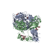
|
|---|---|
| 1 |
|
-Components
-Protein , 2 types, 2 molecules AB
| #1: Protein | Mass: 104460.719 Da / Num. of mol.: 1 / Fragment: ECTODOMAIN, UNP RESIDUES 32-990 / Source method: isolated from a natural source / Details: FITTED FROM PDB ID 3FCS / Source: (natural) Homo sapiens (human) / Tissue: BLOOD / References: UniProt: P08514 |
|---|---|
| #2: Protein | Mass: 76316.945 Da / Num. of mol.: 1 / Fragment: RESIDUES 27-716 / Source method: isolated from a natural source / Details: FITTED FROM PDB ID 3FCS / Source: (natural) Homo sapiens (human) / Cell line: PLATELET / Tissue: BLOOD / References: UniProt: P05106 |
-Sugars , 5 types, 7 molecules 
| #3: Polysaccharide | Source method: isolated from a genetically manipulated source #4: Polysaccharide | alpha-D-mannopyranose-(1-3)-beta-D-mannopyranose-(1-4)-2-acetamido-2-deoxy-beta-D-glucopyranose-(1- ...alpha-D-mannopyranose-(1-3)-beta-D-mannopyranose-(1-4)-2-acetamido-2-deoxy-beta-D-glucopyranose-(1-4)-2-acetamido-2-deoxy-beta-D-glucopyranose | Source method: isolated from a genetically manipulated source #5: Polysaccharide | beta-D-mannopyranose-(1-4)-2-acetamido-2-deoxy-beta-D-glucopyranose-(1-4)-2-acetamido-2-deoxy-beta- ...beta-D-mannopyranose-(1-4)-2-acetamido-2-deoxy-beta-D-glucopyranose-(1-4)-2-acetamido-2-deoxy-beta-D-glucopyranose | Source method: isolated from a genetically manipulated source #6: Polysaccharide | alpha-D-mannopyranose-(1-2)-alpha-D-mannopyranose-(1-3)-beta-D-mannopyranose-(1-4)-2-acetamido-2- ...alpha-D-mannopyranose-(1-2)-alpha-D-mannopyranose-(1-3)-beta-D-mannopyranose-(1-4)-2-acetamido-2-deoxy-beta-D-glucopyranose-(1-4)-2-acetamido-2-deoxy-beta-D-glucopyranose | Source method: isolated from a genetically manipulated source #7: Sugar |  Type: D-saccharide, beta linking / Mass: 221.208 Da / Num. of mol.: 2 Type: D-saccharide, beta linking / Mass: 221.208 Da / Num. of mol.: 2Source method: isolated from a genetically manipulated source Formula: C8H15NO6 |
|---|
-Details
| Has protein modification | Y |
|---|
-Experimental details
-Experiment
| Experiment | Method: ELECTRON MICROSCOPY |
|---|---|
| EM experiment | Aggregation state: PARTICLE / 3D reconstruction method: single particle reconstruction |
- Sample preparation
| Component | Name: INTEGRIN ALPHAIIBBETA3 IN LIPID BILAYER NANODISC / Type: COMPLEX / Details: MICROGRAPHS TAKEN ON CCD |
|---|---|
| Buffer solution | Name: 150 MM NACL, 10 MM HEPES, PH 7.4, 1 MM CACL2 AND 1 MM MGCL2 pH: 7.4 Details: 150 MM NACL, 10 MM HEPES, PH 7.4, 1 MM CACL2 AND 1 MM MGCL2 |
| Specimen | Conc.: 0.02 mg/ml / Embedding applied: NO / Shadowing applied: NO / Staining applied: YES / Vitrification applied: NO |
| EM staining | Type: NEGATIVE / Material: uranyl acetate |
| Specimen support | Details: CARBON |
| Vitrification | Details: VITRIFICATION 1 -- CRYOGEN- NONE, INSTRUMENT- NONE, |
- Electron microscopy imaging
| Experimental equipment |  Model: Tecnai F20 / Image courtesy: FEI Company |
|---|---|
| Microscopy | Model: FEI TECNAI F20 / Date: Feb 1, 2010 Details: LOW DOSE PACKAGE USED. CCD MAGNIFICATION IS 1.76 TIMES FILM MAGNIFICATION |
| Electron gun | Electron source:  FIELD EMISSION GUN / Accelerating voltage: 120 kV / Illumination mode: FLOOD BEAM FIELD EMISSION GUN / Accelerating voltage: 120 kV / Illumination mode: FLOOD BEAM |
| Electron lens | Mode: BRIGHT FIELD / Nominal magnification: 29000 X / Calibrated magnification: 50592 X / Nominal defocus max: 1500 nm / Nominal defocus min: 1200 nm / Cs: 2 mm |
| Specimen holder | Tilt angle max: 50 ° / Tilt angle min: 0 ° |
| Image recording | Electron dose: 13 e/Å2 / Film or detector model: GENERIC TVIPS (4k x 4k) |
| Image scans | Num. digital images: 1500 |
| Radiation wavelength | Relative weight: 1 |
- Processing
| EM software |
| ||||||||||||
|---|---|---|---|---|---|---|---|---|---|---|---|---|---|
| Symmetry | Point symmetry: C1 (asymmetric) | ||||||||||||
| 3D reconstruction | Method: RANDOM CONICAL TILT THEN REFERENCE BASED RECONSTRUCTION Resolution: 20.5 Å / Num. of particles: 25008 / Nominal pixel size: 2.96 Å / Actual pixel size: 2.96 Å / Magnification calibration: 23A LAYERLINE OF TMV Details: INITIAL MODEL FROM RANDOM CONICAL TILT FOLLOWED BY REFERENCE BASED REFINEMENT PDB FILE 3FCS WAS SPLIT INTO 20 SUBDOMAINS. THESE SUBDOMAINS WERE MANUALLY FITTED INTO THE EM VOLUME SUBMISSION ...Details: INITIAL MODEL FROM RANDOM CONICAL TILT FOLLOWED BY REFERENCE BASED REFINEMENT PDB FILE 3FCS WAS SPLIT INTO 20 SUBDOMAINS. THESE SUBDOMAINS WERE MANUALLY FITTED INTO THE EM VOLUME SUBMISSION BASED ON EXPERIMENTAL DATA FROM EMDB EMD-2281. (DEPOSITION ID: 11378). Symmetry type: POINT | ||||||||||||
| Atomic model building | Protocol: RIGID BODY FIT / Space: REAL / Target criteria: Cross-correlation coefficient / Details: METHOD--RIGID BODY REFINEMENT PROTOCOL--X-RAY | ||||||||||||
| Atomic model building | PDB-ID: 3FCS Accession code: 3FCS / Source name: PDB / Type: experimental model | ||||||||||||
| Refinement | Highest resolution: 20.5 Å | ||||||||||||
| Refinement step | Cycle: LAST / Highest resolution: 20.5 Å
|

