 Movie
Movie Controller

[English] 日本語
 Yorodumi
- PDB-4z3k: Human sepiapterin reductase in complex with the cofactor NADP+ an... -
+ Open data

- Basic information
| Entry | Database: PDB / ID: 4z3k | ||||||
|---|---|---|---|---|---|---|---|
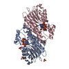
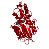
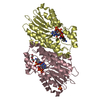
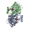
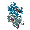
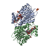
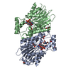
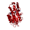
| Title | Human sepiapterin reductase in complex with the cofactor NADP+ and the trypthophan metabolite xanthurenic acid | ||||||
 Components Components | Sepiapterin reductase | ||||||
 Keywords Keywords | OXIDOREDUCTASE / sepiapterin-reductase / xanthurenic acid / inhibitor / tryptophan-metabolite | ||||||
| Function / homology |  Function and homology information Function and homology informationsepiapterin reductase (L-erythro-7,8-dihydrobiopterin-forming) / sepiapterin reductase (NADP+) activity / tetrahydrobiopterin biosynthetic process / alcohol dehydrogenase (NADP+) activity / nitric oxide biosynthetic process / eNOS activation / Tetrahydrobiopterin (BH4) synthesis, recycling, salvage and regulation / NADP binding / mitochondrion / extracellular exosome ...sepiapterin reductase (L-erythro-7,8-dihydrobiopterin-forming) / sepiapterin reductase (NADP+) activity / tetrahydrobiopterin biosynthetic process / alcohol dehydrogenase (NADP+) activity / nitric oxide biosynthetic process / eNOS activation / Tetrahydrobiopterin (BH4) synthesis, recycling, salvage and regulation / NADP binding / mitochondrion / extracellular exosome / nucleoplasm / cytosol Similarity search - Function | ||||||
| Biological species |  Homo sapiens (human) Homo sapiens (human) | ||||||
| Method |  X-RAY DIFFRACTION / SYNCHROTRON / MOLECULAR REPLACEMENT / molecular replacement / Resolution: 2.35 Å X-RAY DIFFRACTION / SYNCHROTRON / MOLECULAR REPLACEMENT / molecular replacement / Resolution: 2.35 Å | ||||||
 Authors Authors | Johnsson, K. | ||||||
 Citation Citation | Journal: J.Biol.Chem. / Year: 2016 Title: Tetrahydrobiopterin Biosynthesis as a Potential Target of the Kynurenine Pathway Metabolite Xanthurenic Acid. Authors: Haruki, H. / Hovius, R. / Pedersen, M.G. / Johnsson, K. | ||||||
| History |
|
- Structure visualization
| Structure viewer | Molecule: MolmilJmol/JSmol |
|---|
- Downloads & links
-Download
| PDBx/mmCIF format | 4z3k.cif.gz | 215.7 KB | Display | PDBx/mmCIF format |
|---|---|---|---|---|
| PDB format | pdb4z3k.ent.gz | 172.7 KB | Display | PDB format |
| PDBx/mmJSON format | 4z3k.json.gz | Tree view | PDBx/mmJSON format | |
| Others |  Other downloads Other downloads |
-Validation report
| Arichive directory | https://data.pdbj.org/pub/pdb/validation_reports/z3/4z3kftp://data.pdbj.org/pub/pdb/validation_reports/z3/4z3k | HTTPS FTP |
|---|
-Related structure data
| Related structure data |  4hwkS S: Starting model for refinement |
|---|---|
| Similar structure data |


-Links
 PDBj
PDBj


- Assembly
| Deposited unit | 
| ||||||||
|---|---|---|---|---|---|---|---|---|---|
| 1 | 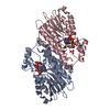
| ||||||||
| 2 | 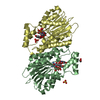
| ||||||||
| Unit cell |
|
-Components
-Protein , 1 types, 4 molecules ABCD
| #1: Protein | Mass: 29891.414 Da / Num. of mol.: 4 Source method: isolated from a genetically manipulated source Details: The N-terminal MHHHHHHENLYFQGMEG is not resolved in the structure & is composed of a His_6 tag; TEV cleavage site; MEG are the 1st 3 residues of the wt protein. The C-terminal K is not visible in the structure. Source: (gene. exp.) Homo sapiens (human) / Gene: SPR / Plasmid: pET9a / Production host:  References: UniProt: P35270, sepiapterin reductase (L-erythro-7,8-dihydrobiopterin-forming) |
|---|
-Non-polymers , 5 types, 175 molecules 


| #2: Chemical | ChemComp-NAP /  Mass: 743.405 Da / Num. of mol.: 4 / Source method: obtained synthetically / Formula: C21H28N7O17P3 Mass: 743.405 Da / Num. of mol.: 4 / Source method: obtained synthetically / Formula: C21H28N7O17P3#3: Chemical | ChemComp-4KL /  Mass: 205.167 Da / Num. of mol.: 4 / Source method: obtained synthetically / Formula: C10H7NO4 / Comment: inhibitor, agonist*YM Mass: 205.167 Da / Num. of mol.: 4 / Source method: obtained synthetically / Formula: C10H7NO4 / Comment: inhibitor, agonist*YM#4: Chemical | ChemComp-SO4 /  Mass: 96.063 Da / Num. of mol.: 6 / Source method: obtained synthetically / Formula: SO4 Mass: 96.063 Da / Num. of mol.: 6 / Source method: obtained synthetically / Formula: SO4#5: Chemical | ChemComp-EDO / |  Mass: 62.068 Da / Num. of mol.: 1 / Source method: obtained synthetically / Formula: C2H6O2 Mass: 62.068 Da / Num. of mol.: 1 / Source method: obtained synthetically / Formula: C2H6O2#6: Water | ChemComp-HOH / | Mass: 18.015 Da / Num. of mol.: 160 / Source method: isolated from a natural source / Formula: H2O |
|---|
-Experimental details
-Experiment
| Experiment | Method: X-RAY DIFFRACTION / Number of used crystals: 1 |
|---|
- Sample preparation
| Crystal | Density Matthews: 4.89 Å3/Da / Density % sol: 74.86 % |
|---|---|
| Crystal grow | Temperature: 277 K / Method: vapor diffusion, hanging drop / pH: 7.5 Details: 2% W/V PEG1000, 1.9 M Ammonium sulfate, 0.1 M Hepes pH7.5 |
-Data collection
| Diffraction | Mean temperature: 100 K | ||||||||||||||||||||||||||||||||||||||||||||||||||||||||||||||||||||||||||||||||||||||||||||||||||||||||||||||
|---|---|---|---|---|---|---|---|---|---|---|---|---|---|---|---|---|---|---|---|---|---|---|---|---|---|---|---|---|---|---|---|---|---|---|---|---|---|---|---|---|---|---|---|---|---|---|---|---|---|---|---|---|---|---|---|---|---|---|---|---|---|---|---|---|---|---|---|---|---|---|---|---|---|---|---|---|---|---|---|---|---|---|---|---|---|---|---|---|---|---|---|---|---|---|---|---|---|---|---|---|---|---|---|---|---|---|---|---|---|---|---|
| Diffraction source | Source: SYNCHROTRON / Site: SLS  / Beamline: X06DA / Wavelength: 1 Å / Beamline: X06DA / Wavelength: 1 Å | ||||||||||||||||||||||||||||||||||||||||||||||||||||||||||||||||||||||||||||||||||||||||||||||||||||||||||||||
| Detector | Type: DECTRIS PILATUS 2M-F / Detector: PIXEL / Date: Nov 4, 2014 | ||||||||||||||||||||||||||||||||||||||||||||||||||||||||||||||||||||||||||||||||||||||||||||||||||||||||||||||
| Radiation | Monochromator: silicon sensor / Protocol: SINGLE WAVELENGTH / Monochromatic (M) / Laue (L): M / Scattering type: x-ray | ||||||||||||||||||||||||||||||||||||||||||||||||||||||||||||||||||||||||||||||||||||||||||||||||||||||||||||||
| Radiation wavelength | Wavelength: 1 Å / Relative weight: 1 | ||||||||||||||||||||||||||||||||||||||||||||||||||||||||||||||||||||||||||||||||||||||||||||||||||||||||||||||
| Reflection | Resolution: 2.35→124.676 Å / Num. all: 88165 / Num. obs: 88165 / % possible obs: 100 % / Redundancy: 12.9 % / Biso Wilson estimate: 57.3 Å2 / Rpim(I) all: 0.031 / Rrim(I) all: 0.111 / Rsym value: 0.106 / Net I/av σ(I): 6.957 / Net I/σ(I): 20 / Num. measured all: 1136272 | ||||||||||||||||||||||||||||||||||||||||||||||||||||||||||||||||||||||||||||||||||||||||||||||||||||||||||||||
| Reflection shell | Diffraction-ID: 1 / Rejects: _
|
-Phasing
| Phasing | Method: molecular replacement | |||||||||
|---|---|---|---|---|---|---|---|---|---|---|
| Phasing MR | Model details: Phaser MODE: MR_AUTO
|
- Processing
| Software |
| |||||||||||||||||||||||||||||||||||||||||||||||||||||||||||||||||||||||||||
|---|---|---|---|---|---|---|---|---|---|---|---|---|---|---|---|---|---|---|---|---|---|---|---|---|---|---|---|---|---|---|---|---|---|---|---|---|---|---|---|---|---|---|---|---|---|---|---|---|---|---|---|---|---|---|---|---|---|---|---|---|---|---|---|---|---|---|---|---|---|---|---|---|---|---|---|---|
| Refinement | Method to determine structure: MOLECULAR REPLACEMENT Starting model: 4HWK Resolution: 2.35→124.676 Å / Cor.coef. Fo:Fc: 0.962 / Cor.coef. Fo:Fc free: 0.946 / WRfactor Rfree: 0.1829 / WRfactor Rwork: 0.1541 / FOM work R set: 0.8359 / SU B: 5.614 / SU ML: 0.127 / SU R Cruickshank DPI: 0.1743 / SU Rfree: 0.1606 / Cross valid method: THROUGHOUT / σ(F): 0 / ESU R: 0.174 / ESU R Free: 0.161 / Stereochemistry target values: MAXIMUM LIKELIHOOD Details: HYDROGENS HAVE BEEN ADDED IN THE RIDING POSITIONS U VALUES : REFINED INDIVIDUALLY
| |||||||||||||||||||||||||||||||||||||||||||||||||||||||||||||||||||||||||||
| Solvent computation | Ion probe radii: 0.8 Å / Shrinkage radii: 0.8 Å / VDW probe radii: 1.2 Å / Solvent model: MASK | |||||||||||||||||||||||||||||||||||||||||||||||||||||||||||||||||||||||||||
| Displacement parameters | Biso max: 146.42 Å2 / Biso mean: 50.093 Å2 / Biso min: 24.72 Å2
| |||||||||||||||||||||||||||||||||||||||||||||||||||||||||||||||||||||||||||
| Refine analyze | Luzzati coordinate error obs: 0.301 Å | |||||||||||||||||||||||||||||||||||||||||||||||||||||||||||||||||||||||||||
| Refinement step | Cycle: final / Resolution: 2.35→124.676 Å
| |||||||||||||||||||||||||||||||||||||||||||||||||||||||||||||||||||||||||||
| Refine LS restraints |
| |||||||||||||||||||||||||||||||||||||||||||||||||||||||||||||||||||||||||||
| LS refinement shell | Resolution: 2.35→2.411 Å / Total num. of bins used: 20
|
