 Movie
Movie Controller

[English] 日本語
 Yorodumi
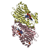
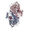
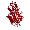
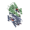
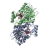
- PDB-4j7x: Crystal structure of human sepiapterin reductase in complex with ... -
+ Open data

- Basic information
| Entry | Database: PDB / ID: 4j7x | ||||||
|---|---|---|---|---|---|---|---|
| Title | Crystal structure of human sepiapterin reductase in complex with sulfasalazine | ||||||
 Components Components | Sepiapterin reductase | ||||||
 Keywords Keywords | OXIDOREDUCTASE / Reductase | ||||||
| Function / homology |  Function and homology information Function and homology informationsepiapterin reductase (L-erythro-7,8-dihydrobiopterin-forming) / sepiapterin reductase (NADP+) activity / tetrahydrobiopterin biosynthetic process / alcohol dehydrogenase (NADP+) activity / nitric oxide biosynthetic process / eNOS activation / Tetrahydrobiopterin (BH4) synthesis, recycling, salvage and regulation / NADP binding / mitochondrion / extracellular exosome ...sepiapterin reductase (L-erythro-7,8-dihydrobiopterin-forming) / sepiapterin reductase (NADP+) activity / tetrahydrobiopterin biosynthetic process / alcohol dehydrogenase (NADP+) activity / nitric oxide biosynthetic process / eNOS activation / Tetrahydrobiopterin (BH4) synthesis, recycling, salvage and regulation / NADP binding / mitochondrion / extracellular exosome / nucleoplasm / cytosol Similarity search - Function | ||||||
| Biological species |  Homo sapiens (human) Homo sapiens (human) | ||||||
| Method |  X-RAY DIFFRACTION / SYNCHROTRON / MOLECULAR REPLACEMENT / Resolution: 2.6 Å X-RAY DIFFRACTION / SYNCHROTRON / MOLECULAR REPLACEMENT / Resolution: 2.6 Å | ||||||
 Authors Authors | Groenlund Pedersen, M. / Pojer, F. / Johnsson, K. | ||||||
 Citation Citation | Journal: To be Published Title: Crystal structure of human sepiapterin reductase in complex with sulfasalazine Authors: Groenlund Pedersen, M. / Pojer, F. / Johnsson, K. | ||||||
| History |
|
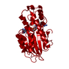
- Structure visualization
| Structure viewer | Molecule: MolmilJmol/JSmol |
|---|
- Downloads & links
-Download
| PDBx/mmCIF format | 4j7x.cif.gz | 215.7 KB | Display | PDBx/mmCIF format |
|---|---|---|---|---|
| PDB format | pdb4j7x.ent.gz | 173.4 KB | Display | PDB format |
| PDBx/mmJSON format | 4j7x.json.gz | Tree view | PDBx/mmJSON format | |
| Others |  Other downloads Other downloads |
-Validation report
| Arichive directory | https://data.pdbj.org/pub/pdb/validation_reports/j7/4j7xftp://data.pdbj.org/pub/pdb/validation_reports/j7/4j7x | HTTPS FTP |
|---|
-Related structure data
| Related structure data |  4hwkS S: Starting model for refinement |
|---|---|
| Similar structure data |


-Links
 PDBj
PDBj


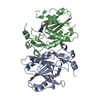
- Assembly
| Deposited unit | 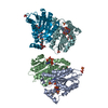
| ||||||||||||
|---|---|---|---|---|---|---|---|---|---|---|---|---|---|
| 1 | 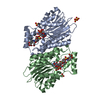
| ||||||||||||
| 2 | 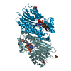
| ||||||||||||
| Unit cell |
| ||||||||||||
| Noncrystallographic symmetry (NCS) | NCS oper:
|
-Components
-Protein , 1 types, 4 molecules ABFJ
| #1: Protein | Mass: 31290.029 Da / Num. of mol.: 4 / Fragment: Full lenght sepiapterin reductase Source method: isolated from a genetically manipulated source Source: (gene. exp.) Homo sapiens (human) / Gene: SPR / Production host:  References: UniProt: P35270, sepiapterin reductase (L-erythro-7,8-dihydrobiopterin-forming) |
|---|
-Non-polymers , 6 types, 130 molecules 

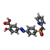


| #2: Chemical | ChemComp-NAP /  Mass: 743.405 Da / Num. of mol.: 4 / Source method: obtained synthetically / Formula: C21H28N7O17P3 Mass: 743.405 Da / Num. of mol.: 4 / Source method: obtained synthetically / Formula: C21H28N7O17P3#3: Chemical | ChemComp-SO4 /  Mass: 96.063 Da / Num. of mol.: 10 / Source method: obtained synthetically / Formula: SO4 Mass: 96.063 Da / Num. of mol.: 10 / Source method: obtained synthetically / Formula: SO4#4: Chemical | ChemComp-SAS /  Mass: 398.393 Da / Num. of mol.: 4 / Source method: obtained synthetically / Formula: C18H14N4O5S / Comment: medication*YM Mass: 398.393 Da / Num. of mol.: 4 / Source method: obtained synthetically / Formula: C18H14N4O5S / Comment: medication*YM#5: Chemical | ChemComp-PEG /  Mass: 106.120 Da / Num. of mol.: 6 / Source method: obtained synthetically / Formula: C4H10O3 Mass: 106.120 Da / Num. of mol.: 6 / Source method: obtained synthetically / Formula: C4H10O3#6: Chemical |  Mass: 92.094 Da / Num. of mol.: 2 / Source method: obtained synthetically / Formula: C3H8O3 Mass: 92.094 Da / Num. of mol.: 2 / Source method: obtained synthetically / Formula: C3H8O3#7: Water | ChemComp-HOH / | Mass: 18.015 Da / Num. of mol.: 104 / Source method: isolated from a natural source / Formula: H2O |
|---|
-Experimental details
-Experiment
| Experiment | Method: X-RAY DIFFRACTION / Number of used crystals: 1 |
|---|
- Sample preparation
| Crystal | Density Matthews: 4.63 Å3/Da / Density % sol: 73.46 % |
|---|---|
| Crystal grow | Temperature: 277 K / Method: vapor diffusion, hanging drop / pH: 7.5 Details: 1.70 M ammonium sulphate, 0.1 M HEPES, 2 % w/v PEG 1000, pH 7.5, VAPOR DIFFUSION, HANGING DROP, temperature 277K |
-Data collection
| Diffraction | Mean temperature: 100 K |
|---|---|
| Diffraction source | Source: SYNCHROTRON / Site: ESRF  / Beamline: BM30A / Wavelength: 1 Å / Beamline: BM30A / Wavelength: 1 Å |
| Detector | Type: ADSC QUANTUM 315r / Detector: CCD / Date: Jun 26, 2011 |
| Radiation | Monochromator: Si(111), Si(311) / Protocol: SINGLE WAVELENGTH / Monochromatic (M) / Laue (L): M / Scattering type: x-ray |
| Radiation wavelength | Wavelength: 1 Å / Relative weight: 1 |
| Reflection | Resolution: 2.6→50 Å / Num. all: 69669 / Num. obs: 68379 / % possible obs: 98.15 % / Observed criterion σ(F): 0 / Observed criterion σ(I): 0 |
| Reflection shell | Resolution: 2.6→2.76 Å / % possible all: 98.3 |
- Processing
| Software |
| |||||||||||||||||||||||||||||||||||||||||||||
|---|---|---|---|---|---|---|---|---|---|---|---|---|---|---|---|---|---|---|---|---|---|---|---|---|---|---|---|---|---|---|---|---|---|---|---|---|---|---|---|---|---|---|---|---|---|---|
| Refinement | Method to determine structure: MOLECULAR REPLACEMENT Starting model: 4HWK Resolution: 2.6→48.84 Å / Cor.coef. Fo:Fc: 0.914 / Cor.coef. Fo:Fc free: 0.9 / SU B: 7.783 / SU ML: 0.163 / Cross valid method: THROUGHOUT / σ(F): 0 / ESU R: 0.292 / ESU R Free: 0.228 / Stereochemistry target values: MAXIMUM LIKELIHOOD / Details: HYDROGENS HAVE BEEN USED IF PRESENT IN THE INPUT
| |||||||||||||||||||||||||||||||||||||||||||||
| Solvent computation | Ion probe radii: 0.8 Å / Shrinkage radii: 0.8 Å / VDW probe radii: 1.2 Å / Solvent model: MASK | |||||||||||||||||||||||||||||||||||||||||||||
| Displacement parameters | Biso mean: 43.517 Å2
| |||||||||||||||||||||||||||||||||||||||||||||
| Refinement step | Cycle: LAST / Resolution: 2.6→48.84 Å
| |||||||||||||||||||||||||||||||||||||||||||||
| Refine LS restraints |
| |||||||||||||||||||||||||||||||||||||||||||||
| LS refinement shell | Resolution: 2.603→2.67 Å / Total num. of bins used: 20
|
