[English] 日本語
Yorodumi
- PDB-4phk: The Structural Basis of Differential Inhibition of Human Calpain ... -

+
Open data


ID or keywords:

Loading...

-
Basic information

Entry
Database: PDB / ID: 4phk
Title
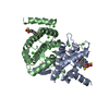
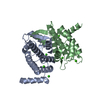
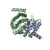
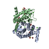
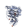
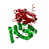
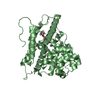
The Structural Basis of Differential Inhibition of Human Calpain by Indole and Phenyl alpha-Mercaptoacrylic Acids. The complex with (Z)-3-(4-chlorophenyl)-2-mercaptoacrylic acid
ComponentsCalpain small subunit 1
KeywordsHYDROLASE / Domain VI / PEF(S) / Calcium Binding / Protease
Function / homology
Function and homology information


calpain complex / calcium-dependent cysteine-type endopeptidase activity / Formation of the cornified envelope / Deregulated CDK5 triggers multiple neurodegenerative pathways in Alzheimer's disease models / regulation of macroautophagy / Degradation of the extracellular matrix / calcium ion binding / positive regulation of cell population proliferation / proteolysis / extracellular exosome ...calpain complex / calcium-dependent cysteine-type endopeptidase activity / Formation of the cornified envelope / Deregulated CDK5 triggers multiple neurodegenerative pathways in Alzheimer's disease models / regulation of macroautophagy / Degradation of the extracellular matrix / calcium ion binding / positive regulation of cell population proliferation / proteolysis / extracellular exosome / membrane / plasma membrane / cytosol
Similarity search - Function
EF-hand / Recoverin; domain 1 / EF-Hand 1, calcium-binding site / EF-hand calcium-binding domain. / EF-hand calcium-binding domain profile. / EF-hand domain / EF-hand domain pair / Orthogonal Bundle / Mainly Alpha
Similarity search - Domain/homology
(Z)-3-(4-chlorophenyl)-2-mercaptoacrylic acid / Calpain small subunit 1
Similarity search - Component
Biological speciesHomo sapiens (human)
MethodX-RAY DIFFRACTION / SYNCHROTRON / Resolution: 2.05 Å
AuthorsRizkallah, P.J. / Allemann, R.K. / Adams, S.E. / Miller, D.J. / Hallett, M.B. / Robinson, E.
Funding support United Kingdom, 1items
OrganizationGrant numberCountry
Medical Research Council (United Kingdom)G0701192 United Kingdom
CitationJournal: J.Struct.Biol. / Year: 2014
Title: The structural basis of differential inhibition of human calpain by indole and phenyl alpha-mercaptoacrylic acids.
Authors: Adams, S.E. / Rizkallah, P.J. / Miller, D.J. / Robinson, E.J. / Hallett, M.B. / Allemann, R.K.
History
DepositionMay 6, 2014Deposition site: RCSB / Processing site: PDBE
Revision 1.0Aug 13, 2014Provider: repository / Type: Initial release
Revision 1.1Sep 17, 2014Group: Database references
Revision 1.2Feb 4, 2015Group: Other
Revision 2.0Sep 13, 2017Group: Atomic model / Author supporting evidence / Derived calculations
Category: atom_site / pdbx_audit_support ...atom_site / pdbx_audit_support / pdbx_struct_conn_angle / struct_conn / struct_site_gen
Item: _atom_site.B_iso_or_equiv / _atom_site.Cartn_x ..._atom_site.B_iso_or_equiv / _atom_site.Cartn_x / _atom_site.Cartn_y / _atom_site.Cartn_z / _pdbx_audit_support.funding_organization / _pdbx_struct_conn_angle.ptnr1_auth_seq_id / _pdbx_struct_conn_angle.ptnr3_auth_seq_id / _struct_conn.ptnr2_auth_seq_id / _struct_site_gen.auth_seq_id
Revision 2.1May 8, 2024Group: Data collection / Database references / Derived calculations
Category: chem_comp_atom / chem_comp_bond ...chem_comp_atom / chem_comp_bond / database_2 / pdbx_struct_conn_angle / struct_conn
Item: _database_2.pdbx_DOI / _database_2.pdbx_database_accession ..._database_2.pdbx_DOI / _database_2.pdbx_database_accession / _pdbx_struct_conn_angle.ptnr1_auth_seq_id / _pdbx_struct_conn_angle.ptnr3_auth_seq_id / _pdbx_struct_conn_angle.value / _struct_conn.pdbx_dist_value / _struct_conn.ptnr1_auth_asym_id / _struct_conn.ptnr1_auth_comp_id / _struct_conn.ptnr1_auth_seq_id / _struct_conn.ptnr1_label_asym_id / _struct_conn.ptnr1_label_atom_id / _struct_conn.ptnr1_label_comp_id / _struct_conn.ptnr1_label_seq_id / _struct_conn.ptnr2_auth_asym_id / _struct_conn.ptnr2_auth_comp_id / _struct_conn.ptnr2_auth_seq_id / _struct_conn.ptnr2_label_asym_id / _struct_conn.ptnr2_label_atom_id / _struct_conn.ptnr2_label_comp_id

-
Structure visualization

Structure viewerMolecule:
MolmilJmol/JSmol

Downloads & links

-
Assembly

Deposited unit
A: Calpain small subunit 1
B: Calpain small subunit 1
hetero molecules


Theoretical massNumber of molelcules
Total (without water)40,78312
Polymers40,0332
Non-polymers75010
Water2,522140
1


  • Idetical with deposited unit
  • defined by software
TypeNameSymmetry operationNumber
identity operation1_555x,y,z1
Buried area4870 Å2
ΔGint-110 kcal/mol
Surface area16140 Å2
MethodPISA
Unit cell
Length a, b, c (Å)49.540, 80.090, 56.890
Angle α, β, γ (deg.)90.00, 91.33, 90.00
Int Tables number4
Space group name H-MP1211

-
Components

#1: Protein Calpain small subunit 1 / CSS1 / Calcium-activated neutral proteinase small subunit / CANP small subunit / Calcium-dependent ...CSS1 / Calcium-activated neutral proteinase small subunit / CANP small subunit / Calcium-dependent protease small subunit / CDPS / Calcium-dependent protease small subunit 1 / Calpain regulatory subunit


Mass: 20016.607 Da / Num. of mol.: 2 / Fragment: residues 96-268
Source method: isolated from a genetically manipulated source
Source: (gene. exp.) Homo sapiens (human) / Gene: CAPNS1, CAPN4, CAPNS / Production host: Escherichia coli (E. coli) / References: UniProt: P04632
#2: Chemical ChemComp-2UB / (Z)-3-(4-chlorophenyl)-2-mercaptoacrylic acid


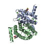
Mass: 214.669 Da / Num. of mol.: 2 / Source method: obtained synthetically / Formula: C9H7ClO2S
#3: Chemical
ChemComp-CA / CALCIUM ION


Mass: 40.078 Da / Num. of mol.: 8 / Source method: obtained synthetically / Formula: Ca
#4: Water ChemComp-HOH / water


Mass: 18.015 Da / Num. of mol.: 140 / Source method: isolated from a natural source / Formula: H2O

-
Experimental details

-
Experiment

ExperimentMethod: X-RAY DIFFRACTION

-
Sample preparation

CrystalDensity Matthews: 2.82 Å3/Da / Density % sol: 56.36 %
Crystal growTemperature: 293 K / Method: vapor diffusion, sitting drop
Details: PROTEIN WAS CRYSTALLIZED FROM 12% PEG6000, 20 MM CACL2, 50 MM CACODYLATE BUFFER, PH7.4

-
Data collection

DiffractionMean temperature: 100 K
Diffraction sourceSource: SYNCHROTRON / Site: Diamond / Beamline: I03 / Wavelength: 0.9673 Å
DetectorType: DECTRIS PILATUS 2M / Detector: PIXEL / Date: Sep 23, 2013
RadiationProtocol: SINGLE WAVELENGTH / Monochromatic (M) / Laue (L): M / Scattering type: x-ray
Radiation wavelengthWavelength: 0.9673 Å / Relative weight: 1
ReflectionResolution: 2.05→80.09 Å / Num. all: 87320 / Num. obs: 25589 / % possible obs: 91.8 % / Redundancy: 3.4 % / Rmerge(I) obs: 0.062 / Net I/σ(I): 10.8
Reflection shellResolution: 2.05→2.1 Å / Redundancy: 2.6 % / Rmerge(I) obs: 0.603 / Mean I/σ(I) obs: 2.3 / % possible all: 70

-
Processing

SoftwareName: REFMAC / Version: 5.8.0049 / Classification: refinement
RefinementResolution: 2.05→80.09 Å / Cor.coef. Fo:Fc: 0.922 / Cor.coef. Fo:Fc free: 0.885 / SU B: 14.24 / SU ML: 0.183 / Cross valid method: THROUGHOUT / ESU R: 0.247 / ESU R Free: 0.212 / Stereochemistry target values: MAXIMUM LIKELIHOOD / Details: HYDROGENS HAVE BEEN ADDED IN THE RIDING POSITIONS
RfactorNum. reflection% reflectionSelection details
Rfree0.27936 1289 5.1 %RANDOM
Rwork0.23073 ---
obs0.23333 24048 90.71 %-
Solvent computationIon probe radii: 0.8 Å / Shrinkage radii: 0.8 Å / VDW probe radii: 1.2 Å / Solvent model: MASK
Displacement parametersBiso mean: 39.244 Å2
Baniso -1Baniso -2Baniso -3
1--0.89 Å20 Å2-1.03 Å2
2--1.24 Å20 Å2
3----0.3 Å2
Refinement stepCycle: 1 / Resolution: 2.05→80.09 Å
ProteinNucleic acidLigandSolventTotal
Num. atoms2806 0 34 140 2980
Refine LS restraints
Refine-IDTypeDev idealDev ideal targetNumber
X-RAY DIFFRACTIONr_bond_refined_d0.0160.0192899
X-RAY DIFFRACTIONr_bond_other_d0.0020.022671
X-RAY DIFFRACTIONr_angle_refined_deg1.7421.9453905
X-RAY DIFFRACTIONr_angle_other_deg0.93336124
X-RAY DIFFRACTIONr_dihedral_angle_1_deg6.1435348
X-RAY DIFFRACTIONr_dihedral_angle_2_deg32.95824.194155
X-RAY DIFFRACTIONr_dihedral_angle_3_deg18.10615514
X-RAY DIFFRACTIONr_dihedral_angle_4_deg13.5751522
X-RAY DIFFRACTIONr_chiral_restr0.1140.2412
X-RAY DIFFRACTIONr_gen_planes_refined0.0080.023341
X-RAY DIFFRACTIONr_gen_planes_other0.0010.02729
X-RAY DIFFRACTIONr_nbd_refined
X-RAY DIFFRACTIONr_nbd_other
X-RAY DIFFRACTIONr_nbtor_refined
X-RAY DIFFRACTIONr_nbtor_other
X-RAY DIFFRACTIONr_xyhbond_nbd_refined
X-RAY DIFFRACTIONr_xyhbond_nbd_other
X-RAY DIFFRACTIONr_metal_ion_refined
X-RAY DIFFRACTIONr_metal_ion_other
X-RAY DIFFRACTIONr_symmetry_vdw_refined
X-RAY DIFFRACTIONr_symmetry_vdw_other
X-RAY DIFFRACTIONr_symmetry_hbond_refined
X-RAY DIFFRACTIONr_symmetry_hbond_other
X-RAY DIFFRACTIONr_symmetry_metal_ion_refined
X-RAY DIFFRACTIONr_symmetry_metal_ion_other
X-RAY DIFFRACTIONr_mcbond_it1.9342.5161389
X-RAY DIFFRACTIONr_mcbond_other1.9332.5151388
X-RAY DIFFRACTIONr_mcangle_it2.8953.7611735
X-RAY DIFFRACTIONr_mcangle_other2.8953.7611736
X-RAY DIFFRACTIONr_scbond_it2.4712.8861510
X-RAY DIFFRACTIONr_scbond_other2.472.8871511
X-RAY DIFFRACTIONr_scangle_it
X-RAY DIFFRACTIONr_scangle_other3.84.2072170
X-RAY DIFFRACTIONr_long_range_B_refined6.64821.613762
X-RAY DIFFRACTIONr_long_range_B_other6.61321.5343739
X-RAY DIFFRACTIONr_rigid_bond_restr
X-RAY DIFFRACTIONr_sphericity_free
X-RAY DIFFRACTIONr_sphericity_bonded
LS refinement shellResolution: 2.05→2.103 Å / Total num. of bins used: 20
RfactorNum. reflection% reflection
Rfree0.613 67 -
Rwork0.598 1263 -
obs--65.32 %
Refinement TLS params.

Method: refined / Refine-ID: X-RAY DIFFRACTION

IDL112)L122)L132)L222)L232)L332)S11 (Å °)S12 (Å °)S13 (Å °)S21 (Å °)S22 (Å °)S23 (Å °)S31 (Å °)S32 (Å °)S33 (Å °)T112)T122)T132)T222)T232)T332)Origin x (Å)Origin y (Å)Origin z (Å)
11.96570.37040.36760.9902-0.18581.19940.1699-0.1921-0.02960.1369-0.07770.16180.1314-0.0695-0.09220.1502-0.03810.01950.0402-0.01240.037513.8092-0.139415.1835
22.1103-0.09520.03030.93620.38730.6811-0.05760.0220.0905-0.12330.11230.0225-0.1320.1014-0.05470.1447-0.0186-0.00070.02220.00820.065928.6779.84570.0649
335.9178-28.1742-19.267826.127717.935712.31360.90721.3140.6013-1.2039-1.55720.9244-0.8381-1.07450.650.21950.0267-0.10480.458-0.04380.579113.0321-11.275922.0782
439.0543-15.553631.45839.7897-10.752326.2207-0.10520.73161.0760.4712-0.9271-0.11070.16410.27711.03230.41-0.06010.1240.1498-0.01770.095139.31585.8764-6.8801
52.32823.7654.80976.18567.475110.98070.0132-0.07940.1361-0.0275-0.18020.2520.10960.05430.1670.09120.01550.010.1132-0.00980.041919.027-3.626316.1339
62.10310.4491.45522.74780.97874.1288-0.02870.1013-0.0826-0.00740.13180.0231-0.03890.062-0.10310.14120.01360.08030.0803-0.01370.111429.84623.504-0.5338
Refinement TLS group
IDRefine-IDRefine TLS-IDAuth asym-IDAuth seq-ID
1X-RAY DIFFRACTION1A96 - 268
2X-RAY DIFFRACTION2B96 - 268
3X-RAY DIFFRACTION3A270
4X-RAY DIFFRACTION4B270
5X-RAY DIFFRACTION5A301 - 304
6X-RAY DIFFRACTION6B301 - 304

+
About Yorodumi

-
News

-
Feb 9, 2022. New format data for meta-information of EMDB entries

New format data for meta-information of EMDB entries

  • Version 3 of the EMDB header file is now the official format.
  • The previous official version 1.9 will be removed from the archive.

Related info.:EMDB header

External links:wwPDB to switch to version 3 of the EMDB data model

-
Aug 12, 2020. Covid-19 info

Covid-19 info

URL: https://pdbjlvh1.pdbj.org/emnavi/covid19.php

New page: Covid-19 featured information page in EM Navigator.

Related info.:Covid-19 info / Mar 5, 2020. Novel coronavirus structure data

+
Mar 5, 2020. Novel coronavirus structure data

Novel coronavirus structure data

Related info.:Yorodumi Speices / Aug 12, 2020. Covid-19 info

External links:COVID-19 featured content - PDBj / Molecule of the Month (242):Coronavirus Proteases

+
Jan 31, 2019. EMDB accession codes are about to change! (news from PDBe EMDB page)

EMDB accession codes are about to change! (news from PDBe EMDB page)

  • The allocation of 4 digits for EMDB accession codes will soon come to an end. Whilst these codes will remain in use, new EMDB accession codes will include an additional digit and will expand incrementally as the available range of codes is exhausted. The current 4-digit format prefixed with “EMD-” (i.e. EMD-XXXX) will advance to a 5-digit format (i.e. EMD-XXXXX), and so on. It is currently estimated that the 4-digit codes will be depleted around Spring 2019, at which point the 5-digit format will come into force.
  • The EM Navigator/Yorodumi systems omit the EMD- prefix.

Related info.:Q: What is EMD? / ID/Accession-code notation in Yorodumi/EM Navigator

External links:EMDB Accession Codes are Changing Soon! / Contact to PDBj

+
Jul 12, 2017. Major update of PDB

Major update of PDB

  • wwPDB released updated PDB data conforming to the new PDBx/mmCIF dictionary.
  • This is a major update changing the version number from 4 to 5, and with Remediation, in which all the entries are updated.
  • In this update, many items about electron microscopy experimental information are reorganized (e.g. em_software).
  • Now, EM Navigator and Yorodumi are based on the updated data.

External links:wwPDB Remediation / Enriched Model Files Conforming to OneDep Data Standards Now Available in the PDB FTP Archive

-
Yorodumi

Thousand views of thousand structures

  • Yorodumi is a browser for structure data from EMDB, PDB, SASBDB, etc.
  • This page is also the successor to EM Navigator detail page, and also detail information page/front-end page for Omokage search.
  • The word "yorodu" (or yorozu) is an old Japanese word meaning "ten thousand". "mi" (miru) is to see.

Related info.:EMDB / PDB / SASBDB / Comparison of 3 databanks / Yorodumi Search / Aug 31, 2016. New EM Navigator & Yorodumi / Yorodumi Papers / Jmol/JSmol / Function and homology information / Changes in new EM Navigator and Yorodumi

Read more