 Movie
Movie Controller

[English] 日本語
 Yorodumi
- PDB-4k6t: Crystal structure of Ad37 fiber knob in complex with trivalent si... -
+ Open data

- Basic information
| Entry | Database: PDB / ID: 4k6t | ||||||
|---|---|---|---|---|---|---|---|
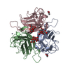
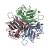
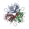
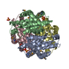
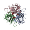
| Title | Crystal structure of Ad37 fiber knob in complex with trivalent sialic acid inhibitor ME0385 | ||||||
 Components Components | Fiber protein | ||||||
 Keywords Keywords | VIRAL PROTEIN / adenovirus / fiber knob / protein carbohydrate interaction / sialic acid / carbohydrate mimic / multivalent ligand | ||||||
| Function / homology |  Function and homology information Function and homology informationadhesion receptor-mediated virion attachment to host cell / viral capsid / cell adhesion / symbiont entry into host cell / host cell nucleus / metal ion binding Similarity search - Function | ||||||
| Biological species |  Human adenovirus 37 Human adenovirus 37 | ||||||
| Method |  X-RAY DIFFRACTION / SYNCHROTRON / MOLECULAR REPLACEMENT / Resolution: 2 Å X-RAY DIFFRACTION / SYNCHROTRON / MOLECULAR REPLACEMENT / Resolution: 2 Å | ||||||
 Authors Authors | Stehle, T. / Bauer, J. | ||||||
 Citation Citation | Journal: Org.Biomol.Chem. / Year: 2015 Title: Triazole linker-based trivalent sialic acid inhibitors of adenovirus type 37 infection of human corneal epithelial cells. Authors: Caraballo, R. / Saleeb, M. / Bauer, J. / Liaci, A.M. / Chandra, N. / Storm, R.J. / Frangsmyr, L. / Qian, W. / Stehle, T. / Arnberg, N. / Elofsson, M. | ||||||
| History |
|
- Structure visualization
| Structure viewer | Molecule: MolmilJmol/JSmol |
|---|
- Downloads & links
-Download
| PDBx/mmCIF format | 4k6t.cif.gz | 464 KB | Display | PDBx/mmCIF format |
|---|---|---|---|---|
| PDB format | pdb4k6t.ent.gz | 382.7 KB | Display | PDB format |
| PDBx/mmJSON format | 4k6t.json.gz | Tree view | PDBx/mmJSON format | |
| Others |  Other downloads Other downloads |
-Validation report
| Arichive directory | https://data.pdbj.org/pub/pdb/validation_reports/k6/4k6tftp://data.pdbj.org/pub/pdb/validation_reports/k6/4k6t | HTTPS FTP |
|---|
-Related structure data


-Links
 PDBj
PDBj

- Assembly
| Deposited unit | 
| ||||||||
|---|---|---|---|---|---|---|---|---|---|
| 1 | 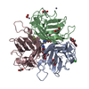
| ||||||||
| 2 | 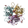
| ||||||||
| Unit cell |
|
-Components
-Protein / Sugars , 2 types, 12 molecules ABCEFG
| #1: Protein | Mass: 21716.535 Da / Num. of mol.: 6 / Fragment: fiber knob Source method: isolated from a genetically manipulated source Source: (gene. exp.) Human adenovirus 37 / Gene: L5 / Plasmid: pPROEX Htb / Production host:  #2: Sugar | ChemComp-SIA /  Type: D-saccharide, alpha linking / Mass: 309.270 Da / Num. of mol.: 6 Type: D-saccharide, alpha linking / Mass: 309.270 Da / Num. of mol.: 6Source method: isolated from a genetically manipulated source Formula: C11H19NO9 |
|---|
-Non-polymers , 9 types, 870 molecules 


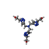


| #3: Chemical | ChemComp-ZN /  Mass: 65.409 Da / Num. of mol.: 14 / Source method: obtained synthetically / Formula: Zn Mass: 65.409 Da / Num. of mol.: 14 / Source method: obtained synthetically / Formula: Zn#4: Chemical | ChemComp-ACT /  Mass: 59.044 Da / Num. of mol.: 9 / Source method: obtained synthetically / Formula: C2H3O2 Mass: 59.044 Da / Num. of mol.: 9 / Source method: obtained synthetically / Formula: C2H3O2#5: Chemical | ChemComp-EDO /  Mass: 62.068 Da / Num. of mol.: 13 / Source method: obtained synthetically / Formula: C2H6O2 Mass: 62.068 Da / Num. of mol.: 13 / Source method: obtained synthetically / Formula: C2H6O2#6: Chemical | ChemComp-CL / |  Mass: 35.453 Da / Num. of mol.: 1 / Source method: obtained synthetically / Formula: Cl Mass: 35.453 Da / Num. of mol.: 1 / Source method: obtained synthetically / Formula: Cl#7: Chemical | ChemComp-1P0 / |  Mass: 392.416 Da / Num. of mol.: 1 / Source method: obtained synthetically / Formula: C15H24N10O3 Mass: 392.416 Da / Num. of mol.: 1 / Source method: obtained synthetically / Formula: C15H24N10O3#8: Chemical | ChemComp-MG /  Mass: 24.305 Da / Num. of mol.: 6 / Source method: obtained synthetically / Formula: Mg Mass: 24.305 Da / Num. of mol.: 6 / Source method: obtained synthetically / Formula: Mg#9: Chemical |  Mass: 92.094 Da / Num. of mol.: 3 / Source method: obtained synthetically / Formula: C3H8O3 Mass: 92.094 Da / Num. of mol.: 3 / Source method: obtained synthetically / Formula: C3H8O3#10: Chemical | ChemComp-CA / |  Mass: 40.078 Da / Num. of mol.: 1 / Source method: obtained synthetically / Formula: Ca Mass: 40.078 Da / Num. of mol.: 1 / Source method: obtained synthetically / Formula: Ca#11: Water | ChemComp-HOH / | Mass: 18.015 Da / Num. of mol.: 822 / Source method: isolated from a natural source / Formula: H2O |
|---|
-Experimental details
-Experiment
| Experiment | Method: X-RAY DIFFRACTION / Number of used crystals: 1 |
|---|
- Sample preparation
| Crystal | Density Matthews: 2.33 Å3/Da / Density % sol: 47.19 % |
|---|---|
| Crystal grow | Temperature: 293 K / Method: vapor diffusion, hanging drop / pH: 6.9 Details: 24% PEG 8000, 0.1M HEPES, 0.05M zinc acetate, pH 6.9, VAPOR DIFFUSION, HANGING DROP, temperature 293K |
-Data collection
| Diffraction | Mean temperature: 100 K |
|---|---|
| Diffraction source | Source: SYNCHROTRON / Site: SLS  / Beamline: X06SA / Wavelength: 1 / Beamline: X06SA / Wavelength: 1 |
| Detector | Type: PSI PILATUS 6M / Detector: PIXEL / Date: Jan 28, 2012 |
| Radiation | Monochromator: Fixed-exit LN2 cooled Double Crystal Monochromator Protocol: SINGLE WAVELENGTH / Monochromatic (M) / Laue (L): M / Scattering type: x-ray |
| Radiation wavelength | Wavelength: 1 Å / Relative weight: 1 |
| Reflection | Resolution: 2→40 Å / Num. all: 80721 / Num. obs: 80719 / % possible obs: 99.3 % / Observed criterion σ(F): -3 / Observed criterion σ(I): -3 / Redundancy: 3 % / Biso Wilson estimate: 30.4 Å2 / Rmerge(I) obs: 0.12 / Net I/σ(I): 6.1 |
| Reflection shell | Resolution: 2→2.04 Å / Redundancy: 3 % / Rmerge(I) obs: 0.563 / Mean I/σ(I) obs: 2.1 / Num. unique all: 5951 / % possible all: 99.1 |
- Processing
| Software |
| |||||||||||||||||||||||||||||||||||||||||||||||||||||||||||||||||||||||||||||||||||||||||||||||||||||||||||||||||||||||||||||||||||||||||||||||||||||||||||||||||||||||||||||||
|---|---|---|---|---|---|---|---|---|---|---|---|---|---|---|---|---|---|---|---|---|---|---|---|---|---|---|---|---|---|---|---|---|---|---|---|---|---|---|---|---|---|---|---|---|---|---|---|---|---|---|---|---|---|---|---|---|---|---|---|---|---|---|---|---|---|---|---|---|---|---|---|---|---|---|---|---|---|---|---|---|---|---|---|---|---|---|---|---|---|---|---|---|---|---|---|---|---|---|---|---|---|---|---|---|---|---|---|---|---|---|---|---|---|---|---|---|---|---|---|---|---|---|---|---|---|---|---|---|---|---|---|---|---|---|---|---|---|---|---|---|---|---|---|---|---|---|---|---|---|---|---|---|---|---|---|---|---|---|---|---|---|---|---|---|---|---|---|---|---|---|---|---|---|---|---|---|
| Refinement | Method to determine structure: MOLECULAR REPLACEMENT Starting model: unliganded Ad37 trimer Resolution: 2→37.26 Å / Cor.coef. Fo:Fc: 0.968 / Cor.coef. Fo:Fc free: 0.951 / SU B: 7.618 / SU ML: 0.11 / Cross valid method: THROUGHOUT / ESU R: 0.161 / ESU R Free: 0.142 / Stereochemistry target values: MAXIMUM LIKELIHOOD / Details: HYDROGENS HAVE BEEN ADDED IN THE RIDING POSITIONS
| |||||||||||||||||||||||||||||||||||||||||||||||||||||||||||||||||||||||||||||||||||||||||||||||||||||||||||||||||||||||||||||||||||||||||||||||||||||||||||||||||||||||||||||||
| Solvent computation | Ion probe radii: 0.8 Å / Shrinkage radii: 0.8 Å / VDW probe radii: 1.2 Å / Solvent model: MASK | |||||||||||||||||||||||||||||||||||||||||||||||||||||||||||||||||||||||||||||||||||||||||||||||||||||||||||||||||||||||||||||||||||||||||||||||||||||||||||||||||||||||||||||||
| Displacement parameters | Biso mean: 26.745 Å2
| |||||||||||||||||||||||||||||||||||||||||||||||||||||||||||||||||||||||||||||||||||||||||||||||||||||||||||||||||||||||||||||||||||||||||||||||||||||||||||||||||||||||||||||||
| Refinement step | Cycle: LAST / Resolution: 2→37.26 Å
| |||||||||||||||||||||||||||||||||||||||||||||||||||||||||||||||||||||||||||||||||||||||||||||||||||||||||||||||||||||||||||||||||||||||||||||||||||||||||||||||||||||||||||||||
| Refine LS restraints |
| |||||||||||||||||||||||||||||||||||||||||||||||||||||||||||||||||||||||||||||||||||||||||||||||||||||||||||||||||||||||||||||||||||||||||||||||||||||||||||||||||||||||||||||||
| LS refinement shell | Resolution: 2→2.052 Å / Total num. of bins used: 20
| |||||||||||||||||||||||||||||||||||||||||||||||||||||||||||||||||||||||||||||||||||||||||||||||||||||||||||||||||||||||||||||||||||||||||||||||||||||||||||||||||||||||||||||||
| Refinement TLS params. | Method: refined / Refine-ID: X-RAY DIFFRACTION
| |||||||||||||||||||||||||||||||||||||||||||||||||||||||||||||||||||||||||||||||||||||||||||||||||||||||||||||||||||||||||||||||||||||||||||||||||||||||||||||||||||||||||||||||
| Refinement TLS group |
|
