Mass: 18.015 Da / Num. of mol.: 370 / Source method: isolated from a natural source / Formula: H2O
-
Details
Compound details
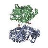
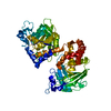
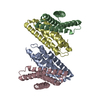
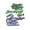
THE PROTEIN CRYSTALLIZED IS THE EXTRACELLULAR LIGAND-BINDING DOMAIN OF GLUA2. TRANSMEMBRANE REGIONS ...THE PROTEIN CRYSTALLIZED IS THE EXTRACELLULAR LIGAND-BINDING DOMAIN OF GLUA2. TRANSMEMBRANE REGIONS WERE GENETICALLY REMOVED AND REPLACED WITH A GLY-THR LINKER (RESIDUES 115-116 IN THE PDB-FILE). THEREFORE THE SEQUENCE MATCHES DISCONTINUOUSLY WITH THE REFERENCE DATABASE (UNP RESIDUES 413-527 AND 653-796, NUMBERING WITH SIGNAL PEPTIDE OF 21 AMINO ACIDS). THE FIRST TWO RESIDUES (GLY, ALA) ARE CLONING REMNANTS
Has protein modification
Y
-
Experimental details
-
Experiment
Experiment
Method: X-RAY DIFFRACTION / Number of used crystals: 1
-
Sample preparation
Crystal
Density Matthews: 2.44 Å3/Da / Density % sol: 49.56 %
Crystal grow
Temperature: 280 K / Method: vapor diffusion, hanging drop / pH: 4.5 Details: 24.4% PEG4000, 0.3M lithium sulfate, 0.1M phosphate-citrate, pH 4.5, VAPOR DIFFUSION, HANGING DROP, temperature 280K
-
Data collection
Diffraction
Mean temperature: 100 K
Diffraction source
Source: SYNCHROTRON / Site: MAX II / Beamline: I911-3 / Wavelength: 1 Å
Detector
Type: MARMOSAIC 225 mm CCD / Detector: CCD / Date: Jun 24, 2011
Radiation
Protocol: SINGLE WAVELENGTH / Monochromatic (M) / Laue (L): M / Scattering type: x-ray
Radiation wavelength
Wavelength: 1 Å / Relative weight: 1
Reflection
Resolution: 2.05→29.44 Å / Num. obs: 36483 / % possible obs: 99.5 % / Redundancy: 5.5 % / Biso Wilson estimate: 21.2 Å2 / Rmerge(I) obs: 0.09 / Rsym value: 0.09 / Net I/σ(I): 7.5
In the structure databanks used in Yorodumi, some data are registered as the other names, "COVID-19 virus" and "2019-nCoV". Here are the details of the virus and the list of structure data.
Jan 31, 2019. EMDB accession codes are about to change! (news from PDBe EMDB page)
EMDB accession codes are about to change! (news from PDBe EMDB page)
The allocation of 4 digits for EMDB accession codes will soon come to an end. Whilst these codes will remain in use, new EMDB accession codes will include an additional digit and will expand incrementally as the available range of codes is exhausted. The current 4-digit format prefixed with “EMD-” (i.e. EMD-XXXX) will advance to a 5-digit format (i.e. EMD-XXXXX), and so on. It is currently estimated that the 4-digit codes will be depleted around Spring 2019, at which point the 5-digit format will come into force.
The EM Navigator/Yorodumi systems omit the EMD- prefix.
Related info.:Q: What is EMD? / ID/Accession-code notation in Yorodumi/EM Navigator
Yorodumi is a browser for structure data from EMDB, PDB, SASBDB, etc.
This page is also the successor to EM Navigator detail page, and also detail information page/front-end page for Omokage search.
The word "yorodu" (or yorozu) is an old Japanese word meaning "ten thousand". "mi" (miru) is to see.
Related info.:EMDB / PDB / SASBDB / Comparison of 3 databanks / Yorodumi Search / Aug 31, 2016. New EM Navigator & Yorodumi / Yorodumi Papers / Jmol/JSmol / Function and homology information / Changes in new EM Navigator and Yorodumi
 Movie
Movie Controller

 Yorodumi
 Open data
 Basic information
 Components
 Keywords
 Function and homology information
 X-RAY DIFFRACTION /
  Authors
 Citation
 Structure visualization
 Downloads & links
 Other downloads


 PDBj
PDBj


 Assembly
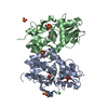


 Type: L-peptide linking / Mass: 173.167 Da / Num. of mol.: 2 / Source method: obtained synthetically / Formula: C7H11NO4
 Mass: 96.063 Da / Num. of mol.: 7 / Source method: obtained synthetically / Formula: SO4
 Mass: 35.453 Da / Num. of mol.: 3 / Source method: obtained synthetically / Formula: Cl
 Mass: 92.094 Da / Num. of mol.: 2 / Source method: obtained synthetically / Formula: C3H8O3
 Sample preparation
 / Beamline: I911-3 / Wavelength: 1 Å
 Processing