 Movie
Movie Controller


+ Open data

- Basic information
| Entry | Database: PDB / ID: 4ask | ||||||
|---|---|---|---|---|---|---|---|
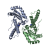
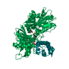
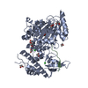
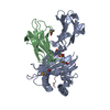
| Title | CRYSTAL STRUCTURE OF JMJD3 WITH GSK-J1 | ||||||
 Components Components | LYSINE-SPECIFIC DEMETHYLASE 6B | ||||||
 Keywords Keywords | OXIDOREDUCTASE / KDM6B / GSK-J1 / INHIBITOR / LYSINE SPECIFIC HISTONE DEMETHYLASE | ||||||
| Function / homology |  Function and homology information Function and homology information[histone H3]-trimethyl-L-lysine27 demethylase / histone H3K27me2/H3K27me3 demethylase activity / endothelial cell differentiation / MLL3/4 complex / inflammatory response to antigenic stimulus / cardiac muscle cell differentiation / mesodermal cell differentiation / histone demethylase activity / cell fate commitment / response to fungicide ...[histone H3]-trimethyl-L-lysine27 demethylase / histone H3K27me2/H3K27me3 demethylase activity / endothelial cell differentiation / MLL3/4 complex / inflammatory response to antigenic stimulus / cardiac muscle cell differentiation / mesodermal cell differentiation / histone demethylase activity / cell fate commitment / response to fungicide / response to activity / Chromatin modifications during the maternal to zygotic transition (MZT) / hippocampus development / HDMs demethylate histones / chromatin DNA binding / beta-catenin binding / cellular response to hydrogen peroxide / positive regulation of cold-induced thermogenesis / heart development / regulation of gene expression / Oxidative Stress Induced Senescence / RNA polymerase II cis-regulatory region sequence-specific DNA binding / chromatin remodeling / positive regulation of transcription by RNA polymerase II / nucleoplasm / metal ion binding / nucleus Similarity search - Function | ||||||
| Biological species |  HOMO SAPIENS (human) HOMO SAPIENS (human) | ||||||
| Method |  X-RAY DIFFRACTION / SYNCHROTRON / MOLECULAR REPLACEMENT / Resolution: 1.86 Å X-RAY DIFFRACTION / SYNCHROTRON / MOLECULAR REPLACEMENT / Resolution: 1.86 Å | ||||||
 Authors Authors | Chung, C. / Mosley, J. / Liddle, J. | ||||||
 Citation Citation | Journal: Nature / Year: 2012 Title: A Selective Jumonji H3K27 Demethylase Inhibitor Modulates the Proinflammatory Macrophage Response Authors: Kruidenier, L. / Chung, C. / Cheng, Z. / Liddle, J. / Che, K. / Joberty, G. / Bantscheff, M. / Bountra, C. / Bridges, A. / Diallo, H. / Eberhard, D. / Hutchinson, S. / Jones, E. / Katso, R. ...Authors: Kruidenier, L. / Chung, C. / Cheng, Z. / Liddle, J. / Che, K. / Joberty, G. / Bantscheff, M. / Bountra, C. / Bridges, A. / Diallo, H. / Eberhard, D. / Hutchinson, S. / Jones, E. / Katso, R. / Leveridge, M. / Mander, P.K. / Mosley, J. / Ramirez-Molina, C. / Rowland, P. / Schofield, C.J. / Sheppard, R.J. / Smith, J.E. / Swales, C. / Tanner, R. / Thomas, P. / Tumber, A. / Drewes, G. / Oppermann, U. / Patel, D.J. / Lee, K. / Wilson, D.M. | ||||||
| History |
|
- Structure visualization
| Structure viewer | Molecule: MolmilJmol/JSmol |
|---|
- Downloads & links
-Download
| PDBx/mmCIF format | 4ask.cif.gz | 377.1 KB | Display | PDBx/mmCIF format |
|---|---|---|---|---|
| PDB format | pdb4ask.ent.gz | 305.8 KB | Display | PDB format |
| PDBx/mmJSON format | 4ask.json.gz | Tree view | PDBx/mmJSON format | |
| Others |  Other downloads Other downloads |
-Validation report
| Arichive directory | https://data.pdbj.org/pub/pdb/validation_reports/as/4askftp://data.pdbj.org/pub/pdb/validation_reports/as/4ask | HTTPS FTP |
|---|
-Related structure data
| Related structure data |  2xueSC  4eyuC  4ez4C  4ezhC S: Starting model for refinement C: citing same article ( |
|---|---|
| Similar structure data |


-Links
 PDBj
PDBj

- Assembly
| Deposited unit | 
| ||||||||
|---|---|---|---|---|---|---|---|---|---|
| 1 | 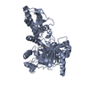
| ||||||||
| 2 | 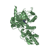
| ||||||||
| Unit cell |
|
-Components
| #1: Protein | Mass: 58050.285 Da / Num. of mol.: 2 / Fragment: CATALYTIC AND ZBD DOMAIN Source method: isolated from a genetically manipulated source Source: (gene. exp.) HOMO SAPIENS (human) / Cell line (production host): SF9 / Production host:   SPODOPTERA FRUGIPERDA (fall armyworm) SPODOPTERA FRUGIPERDA (fall armyworm)References: UniProt: O15054, Oxidoreductases; Acting on paired donors, with incorporation or reduction of molecular oxygen; With 2-oxoglutarate as one donor, and incorporation of one atom of oxygen into each donor #2: Chemical |   Mass: 65.409 Da / Num. of mol.: 2 / Source method: obtained synthetically / Formula: Zn Mass: 65.409 Da / Num. of mol.: 2 / Source method: obtained synthetically / Formula: Zn#3: Chemical |   Mass: 389.450 Da / Num. of mol.: 2 / Source method: obtained synthetically / Formula: C22H23N5O2 Mass: 389.450 Da / Num. of mol.: 2 / Source method: obtained synthetically / Formula: C22H23N5O2#4: Chemical |   Mass: 58.933 Da / Num. of mol.: 2 / Source method: obtained synthetically / Formula: Co Mass: 58.933 Da / Num. of mol.: 2 / Source method: obtained synthetically / Formula: Co#5: Water | ChemComp-HOH / |  Mass: 18.015 Da / Num. of mol.: 1239 / Source method: isolated from a natural source / Formula: H2O Mass: 18.015 Da / Num. of mol.: 1239 / Source method: isolated from a natural source / Formula: H2O |
|---|
-Experimental details
-Experiment
| Experiment | Method: X-RAY DIFFRACTION / Number of used crystals: 1 |
|---|
- Sample preparation
| Crystal | Density Matthews: 2.3 Å3/Da / Density % sol: 46.5 % / Description: NONE |
|---|---|
| Crystal grow | pH: 8.5 Details: 0.1M TRIS HCL, BICINE PH 8.5, 37.5% MPD, PEG1K, PEG3350, 0.03M MGCL2, 0.03M CACL2 |
-Data collection
| Diffraction | Mean temperature: 100 K |
|---|---|
| Diffraction source | Source: SYNCHROTRON / Site: Diamond  / Beamline: I04 / Wavelength: 0.97625 / Beamline: I04 / Wavelength: 0.97625 |
| Detector | Type: ADSC CCD / Detector: CCD / Date: Sep 29, 2010 |
| Radiation | Protocol: SINGLE WAVELENGTH / Monochromatic (M) / Laue (L): M / Scattering type: x-ray |
| Radiation wavelength | Wavelength: 0.97625 Å / Relative weight: 1 |
| Reflection | Resolution: 1.86→40 Å / Num. obs: 83457 / % possible obs: 96.3 % / Observed criterion σ(I): 2 / Redundancy: 1.9 % / Rmerge(I) obs: 0.07 / Net I/σ(I): 12.5 |
| Reflection shell | Resolution: 1.86→1.89 Å / Redundancy: 1.8 % / Rmerge(I) obs: 0.36 / Mean I/σ(I) obs: 1.9 / % possible all: 93.4 |
- Processing
| Software |
| ||||||||||||||||||||||||||||||||||||||||||||||||||||||||||||||||||||||||||||||||||||||||||||||||||||||||||||||||||||||||||||||||||||||||||||||||||||||||||||||||||||||||||||||||||||||
|---|---|---|---|---|---|---|---|---|---|---|---|---|---|---|---|---|---|---|---|---|---|---|---|---|---|---|---|---|---|---|---|---|---|---|---|---|---|---|---|---|---|---|---|---|---|---|---|---|---|---|---|---|---|---|---|---|---|---|---|---|---|---|---|---|---|---|---|---|---|---|---|---|---|---|---|---|---|---|---|---|---|---|---|---|---|---|---|---|---|---|---|---|---|---|---|---|---|---|---|---|---|---|---|---|---|---|---|---|---|---|---|---|---|---|---|---|---|---|---|---|---|---|---|---|---|---|---|---|---|---|---|---|---|---|---|---|---|---|---|---|---|---|---|---|---|---|---|---|---|---|---|---|---|---|---|---|---|---|---|---|---|---|---|---|---|---|---|---|---|---|---|---|---|---|---|---|---|---|---|---|---|---|---|
| Refinement | Method to determine structure: MOLECULAR REPLACEMENT Starting model: PDB ENTRY 2XUE Resolution: 1.86→71.37 Å / Cor.coef. Fo:Fc: 0.964 / Cor.coef. Fo:Fc free: 0.94 / SU B: 5.442 / SU ML: 0.087 / Cross valid method: THROUGHOUT / ESU R: 0.131 / ESU R Free: 0.128 / Stereochemistry target values: MAXIMUM LIKELIHOOD / Details: HYDROGENS HAVE BEEN ADDED IN THE RIDING POSITIONS.
| ||||||||||||||||||||||||||||||||||||||||||||||||||||||||||||||||||||||||||||||||||||||||||||||||||||||||||||||||||||||||||||||||||||||||||||||||||||||||||||||||||||||||||||||||||||||
| Solvent computation | Ion probe radii: 0.8 Å / Shrinkage radii: 0.8 Å / VDW probe radii: 1.2 Å / Solvent model: BABINET MODEL WITH MASK | ||||||||||||||||||||||||||||||||||||||||||||||||||||||||||||||||||||||||||||||||||||||||||||||||||||||||||||||||||||||||||||||||||||||||||||||||||||||||||||||||||||||||||||||||||||||
| Displacement parameters | Biso mean: 23.283 Å2
| ||||||||||||||||||||||||||||||||||||||||||||||||||||||||||||||||||||||||||||||||||||||||||||||||||||||||||||||||||||||||||||||||||||||||||||||||||||||||||||||||||||||||||||||||||||||
| Refinement step | Cycle: LAST / Resolution: 1.86→71.37 Å
| ||||||||||||||||||||||||||||||||||||||||||||||||||||||||||||||||||||||||||||||||||||||||||||||||||||||||||||||||||||||||||||||||||||||||||||||||||||||||||||||||||||||||||||||||||||||
| Refine LS restraints |
|