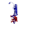
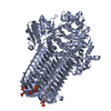
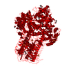
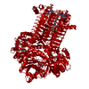
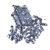
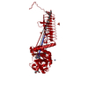
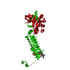
Entry Database : PDB / ID : 4ac3Title S.pneumoniae GlmU in complex with an antibacterial inhibitor BIFUNCTIONAL PROTEIN GLMU Keywords / / Function / homology Function Domain/homology Component
/ / / / / / / / / / / / / / / / / / / / / / / / / / / / / / / / / / / / / / / / / / / / / / / / / Biological species STREPTOCOCCUS PNEUMONIAE (bacteria)Method / / Resolution : 2.1 Å Authors Otterbein, L. / Breed, J. / Ogg, D.J. Journal : Bioorg.Med.Chem.Lett. / Year : 2012Title : Inhibitors of Acetyltransferase Domain of N-Acetylglucosamine-1-Phosphate-Uridyltransferase/ Glucosamine-1-Phosphate-Acetyltransferase (Glmu). Part 1: Hit to Lead Evaluation of a Novel Arylsulfonamide Series.Authors : Green, O.M. / McKenzie, A.R. / Shapiro, A.B. / Otterbein, L. / Ni, H. / Patten, A. / Stokes, S. / Albert, R. / Kawatkar, S. / Breed, J. History Deposition Dec 12, 2011 Deposition site / Processing site Revision 1.0 Feb 15, 2012 Provider / Type Revision 1.1 Aug 15, 2012 Group Revision 1.2 Dec 27, 2017 Group / Category / Item Revision 1.3 Dec 20, 2023 Group Data collection / Database references ... Data collection / Database references / Derived calculations / Other / Refinement description Category chem_comp_atom / chem_comp_bond ... chem_comp_atom / chem_comp_bond / database_2 / pdbx_database_status / pdbx_initial_refinement_model / struct_site Item _database_2.pdbx_DOI / _database_2.pdbx_database_accession ... _database_2.pdbx_DOI / _database_2.pdbx_database_accession / _pdbx_database_status.status_code_sf / _struct_site.pdbx_auth_asym_id / _struct_site.pdbx_auth_comp_id / _struct_site.pdbx_auth_seq_id
Show all Show less
 Movie
Movie Controller


 Open data
 Basic information
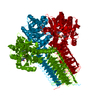
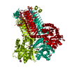
 Components
 Keywords
 Function and homology information
 STREPTOCOCCUS PNEUMONIAE (bacteria)
 X-RAY DIFFRACTION /
  Authors
 Citation
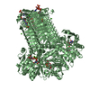
 Structure visualization
 Downloads & links
 Other downloads
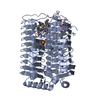

 PDBj
PDBj


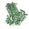
 Assembly
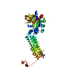

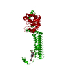
 Mass: 447.548 Da / Num. of mol.: 1 / Source method: obtained synthetically / Formula: C22H29N3O5S
 Mass: 96.063 Da / Num. of mol.: 1 / Source method: obtained synthetically / Formula: SO4
 Mass: 18.015 Da / Num. of mol.: 372 / Source method: isolated from a natural source / Formula: H2O
 Sample preparation
 Processing