 Movie
Movie Controller


+ Open data

- Basic information
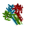
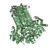
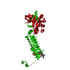
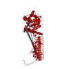
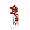
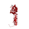
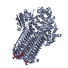
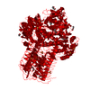
| Entry | Database: PDB / ID: 1g95 | ||||||
|---|---|---|---|---|---|---|---|
| Title | CRYSTAL STRUCTURE OF S.PNEUMONIAE GLMU, APO FORM | ||||||
 Components Components | N-ACETYLGLUCOSAMINE-1-PHOSPHATE URIDYLTRANSFERASE | ||||||
 Keywords Keywords | TRANSFERASE / GlmU / Acetyltransferase / uridyltransferase pyrophosphorylase / left-handed beta-sheet helix / trimer | ||||||
| Function / homology |  Function and homology information Function and homology informationglucosamine-1-phosphate N-acetyltransferase / glucosamine-1-phosphate N-acetyltransferase activity / UDP-N-acetylglucosamine diphosphorylase / UDP-N-acetylglucosamine diphosphorylase activity / UDP-N-acetylglucosamine biosynthetic process / lipid A biosynthetic process / peptidoglycan biosynthetic process / cell wall organization / cell morphogenesis / regulation of cell shape ...glucosamine-1-phosphate N-acetyltransferase / glucosamine-1-phosphate N-acetyltransferase activity / UDP-N-acetylglucosamine diphosphorylase / UDP-N-acetylglucosamine diphosphorylase activity / UDP-N-acetylglucosamine biosynthetic process / lipid A biosynthetic process / peptidoglycan biosynthetic process / cell wall organization / cell morphogenesis / regulation of cell shape / magnesium ion binding / membrane / cytoplasm Similarity search - Function | ||||||
| Biological species |   Streptococcus pneumoniae (bacteria) Streptococcus pneumoniae (bacteria) | ||||||
| Method |  X-RAY DIFFRACTION / SYNCHROTRON / SIRAS / Resolution: 2.33 Å X-RAY DIFFRACTION / SYNCHROTRON / SIRAS / Resolution: 2.33 Å | ||||||
 Authors Authors | Kostrewa, D. / D'Arcy, A. / Kamber, M. | ||||||
 Citation Citation | Journal: J.Mol.Biol. / Year: 2001 Title: Crystal structures of Streptococcus pneumoniae N-acetylglucosamine-1-phosphate uridyltransferase, GlmU, in apo form at 2.33 A resolution and in complex with UDP-N-acetylglucosamine and Mg(2+) at 1.96 A resolution. Authors: Kostrewa, D. / D'Arcy, A. / Takacs, B. / Kamber, M. | ||||||
| History |
|
- Structure visualization
| Structure viewer | Molecule: MolmilJmol/JSmol |
|---|
- Downloads & links
-Download
| PDBx/mmCIF format | 1g95.cif.gz | 101.7 KB | Display | PDBx/mmCIF format |
|---|---|---|---|---|
| PDB format | pdb1g95.ent.gz | 78.6 KB | Display | PDB format |
| PDBx/mmJSON format | 1g95.json.gz | Tree view | PDBx/mmJSON format | |
| Others |  Other downloads Other downloads |
-Validation report
| Arichive directory | https://data.pdbj.org/pub/pdb/validation_reports/g9/1g95ftp://data.pdbj.org/pub/pdb/validation_reports/g9/1g95 | HTTPS FTP |
|---|
-Related structure data

-Links
 PDBj
PDBj


- Assembly
| Deposited unit | 
| ||||||||
|---|---|---|---|---|---|---|---|---|---|
| 1 | 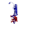
| ||||||||
| Unit cell |
| ||||||||
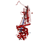
| Details | The biological assembly is a trimer generated from the monomer in the asymmetric unit by the operations: -Y+1, X-Y, Z and -X+Y+1, -X+1, Z |
-Components
| #1: Protein | Mass: 49398.387 Da / Num. of mol.: 1 Source method: isolated from a genetically manipulated source Source: (gene. exp.) Streptococcus pneumoniae (bacteria) / Production host: References: UniProt: Q97R46, UDP-N-acetylglucosamine diphosphorylase |
|---|---|
| #2: Water | ChemComp-HOH /  Mass: 18.015 Da / Num. of mol.: 276 / Source method: isolated from a natural source / Formula: H2O Mass: 18.015 Da / Num. of mol.: 276 / Source method: isolated from a natural source / Formula: H2O |
-Experimental details
-Experiment
| Experiment | Method: X-RAY DIFFRACTION / Number of used crystals: 1 |
|---|
- Sample preparation
| Crystal | Density Matthews: 2.46 Å3/Da / Density % sol: 50 % | |||||||||||||||||||||||||
|---|---|---|---|---|---|---|---|---|---|---|---|---|---|---|---|---|---|---|---|---|---|---|---|---|---|---|
| Crystal grow | Temperature: 298 K / Method: vapor diffusion, hanging drop / pH: 6.5 Details: 0.1 M BIS/TRIS, 0.2 M ammonium sulfate, 25 % PEG 3350, pH 6.5, VAPOR DIFFUSION, HANGING DROP, temperature 298K | |||||||||||||||||||||||||
| Crystal grow | *PLUS Method: vapor diffusion | |||||||||||||||||||||||||
| Components of the solutions | *PLUS
|
-Data collection
| Diffraction | Mean temperature: 120 K |
|---|---|
| Diffraction source | Source: SYNCHROTRON / Site: ESRF  / Beamline: BM1A / Wavelength: 0.873 / Wavelength: 0.873 Å / Beamline: BM1A / Wavelength: 0.873 / Wavelength: 0.873 Å |
| Detector | Type: MARRESEARCH / Detector: IMAGE PLATE / Date: Apr 26, 1998 / Details: Mirrors |
| Radiation | Monochromator: SAGITALLY FOCUSED Si(111) / Protocol: SINGLE WAVELENGTH / Monochromatic (M) / Laue (L): M / Scattering type: x-ray |
| Radiation wavelength | Wavelength: 0.873 Å / Relative weight: 1 |
| Reflection | Resolution: 2.33→30 Å / Num. all: 37195 / Num. obs: 20586 / % possible obs: 97 % / Observed criterion σ(F): 0 / Observed criterion σ(I): 0 / Redundancy: 1.8 % / Biso Wilson estimate: 37.3 Å2 / Rsym value: 0.061 / Net I/σ(I): 10.5 |
| Reflection shell | Resolution: 2.33→2.41 Å / Redundancy: 1.7 % / Mean I/σ(I) obs: 3.1 / Num. unique all: 2007 / Rsym value: 0.188 / % possible all: 95.7 |
| Reflection | *PLUS Rmerge(I) obs: 0.061 |
| Reflection shell | *PLUS % possible obs: 95.7 % / Rmerge(I) obs: 0.188 |
- Processing
| Software |
| ||||||||||||||||||||||||||||||||||||
|---|---|---|---|---|---|---|---|---|---|---|---|---|---|---|---|---|---|---|---|---|---|---|---|---|---|---|---|---|---|---|---|---|---|---|---|---|---|
| Refinement | Method to determine structure: SIRAS / Resolution: 2.33→30 Å / Isotropic thermal model: isotropic / Cross valid method: FREE-R / σ(F): 0 / σ(I): 0 / Stereochemistry target values: Engh & Huber Details: BULK SOLVENT CORRECTION WITH ELECTRON DENSITY = 0.26 E/A**3, B-FACTOR = 23.5 A**2. THE FOLLOWING AMINO ACID RESIDUES WERE NOT VISIBLE IN THE ELECTRON DENSITY MAPS: 188-193, 453-459. THE ...Details: BULK SOLVENT CORRECTION WITH ELECTRON DENSITY = 0.26 E/A**3, B-FACTOR = 23.5 A**2. THE FOLLOWING AMINO ACID RESIDUES WERE NOT VISIBLE IN THE ELECTRON DENSITY MAPS: 188-193, 453-459. THE ELECTRON DENSITY MAPS OF THE FOLLOWING AMINO ACID RESIDUES WERE OF RELATIVELY POOR QUALITY: 2, 10-12, 51-94, 145-149, 154-164, 179-187, 194-197, 439-441, 447-452. THE WATER MOLECULES ARE ORDERED WITH ASCENDING B-FACTORS.
| ||||||||||||||||||||||||||||||||||||
| Solvent computation | Solvent model: bulk solvent mask / Bsol: 23.5 Å2 / ksol: 0.26 e/Å3 | ||||||||||||||||||||||||||||||||||||
| Displacement parameters | Biso mean: 49.1 Å2
| ||||||||||||||||||||||||||||||||||||
| Refine analyze |
| ||||||||||||||||||||||||||||||||||||
| Refinement step | Cycle: LAST / Resolution: 2.33→30 Å
| ||||||||||||||||||||||||||||||||||||
| Refine LS restraints |
| ||||||||||||||||||||||||||||||||||||
| LS refinement shell | Resolution: 2.33→2.41 Å / Total num. of bins used: 10
| ||||||||||||||||||||||||||||||||||||
| Xplor file |
| ||||||||||||||||||||||||||||||||||||
| Software | *PLUS Name: CNS / Classification: refinement | ||||||||||||||||||||||||||||||||||||
| Refinement | *PLUS Lowest resolution: 30 Å / σ(F): 0 / % reflection Rfree: 5 % / Rfactor obs: 0.23 | ||||||||||||||||||||||||||||||||||||
| Solvent computation | *PLUS | ||||||||||||||||||||||||||||||||||||
| Displacement parameters | *PLUS Biso mean: 49.1 Å2 | ||||||||||||||||||||||||||||||||||||
| LS refinement shell | *PLUS Rfactor Rfree: 0.35 / % reflection Rfree: 5 % / Rfactor Rwork: 0.306 |
