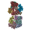
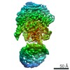
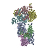
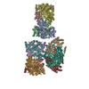
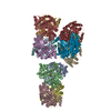
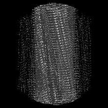
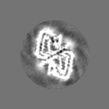
ジャーナル: Nat Commun / 年: 2019 タイトル: Atomic structure of PI3-kinase SH3 amyloid fibrils by cryo-electron microscopy. 著者: Christine Röder / Nicola Vettore / Lena N Mangels / Lothar Gremer / Raimond B G Ravelli / Dieter Willbold / Wolfgang Hoyer / Alexander K Buell / Gunnar F Schröder / 要旨: High resolution structural information on amyloid fibrils is crucial for the understanding of their formation mechanisms and for the rational design of amyloid inhibitors in the context of protein ...High resolution structural information on amyloid fibrils is crucial for the understanding of their formation mechanisms and for the rational design of amyloid inhibitors in the context of protein misfolding diseases. The Src-homology 3 domain of phosphatidyl-inositol-3-kinase (PI3K-SH3) is a model amyloid system that plays a pivotal role in our basic understanding of protein misfolding and aggregation. Here, we present the atomic model of the PI3K-SH3 amyloid fibril with a resolution determined to 3.4 Å by cryo-electron microscopy (cryo-EM). The fibril is composed of two intertwined protofilaments that create an interface spanning 13 residues from each monomer. The model comprises residues 1-77 out of 86 amino acids in total, with the missing residues located in the highly flexible C-terminus. The fibril structure allows us to rationalise the effects of chemically conservative point mutations as well as of the previously reported sequence perturbations on PI3K-SH3 fibril formation and growth.
照射モード: FLOOD BEAM / 撮影モード: BRIGHT FIELD / Cs: 2.7 mm
実験機器
モデル: Talos Arctica / 画像提供: FEI Company
+
画像解析
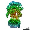
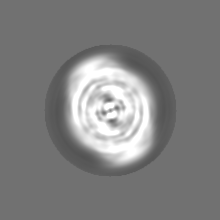
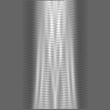
最終 再構成
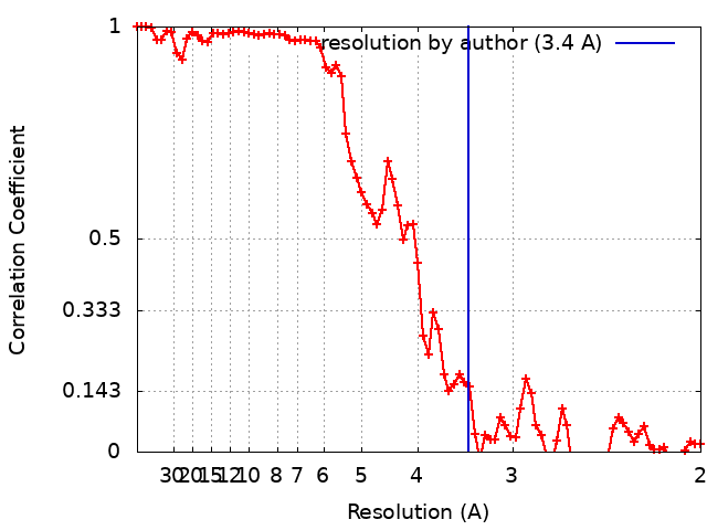
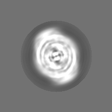
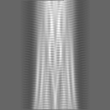
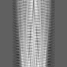
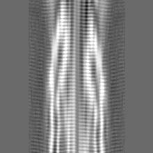
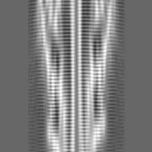
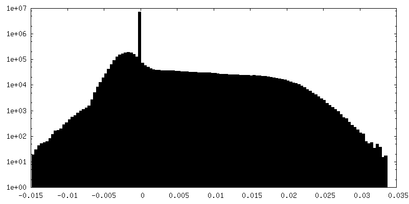
想定した対称性 - らせんパラメータ - Δz: 2.3548 Å 想定した対称性 - らせんパラメータ - ΔΦ: 179.436 ° 想定した対称性 - らせんパラメータ - 軸対称性: C1 (非対称) 解像度のタイプ: BY AUTHOR / 解像度: 3.4 Å / 解像度の算出法: FSC 0.143 CUT-OFF / ソフトウェア - 名称: RELION (ver. 2) 詳細: For the even/odd test, the segment images were split by entire fibrils and refined separately for 25 iterations using the same reference density (low-passed filtered to 20 A). 使用した粒子像数: 27681
Segment selection
選択した数: 103733 / 詳細: Filaments were picked manually in Relion2
初期モデル
モデルのタイプ: NONE
最終 角度割当
タイプ: NOT APPLICABLE / ソフトウェア - 名称: RELION (ver. 2)
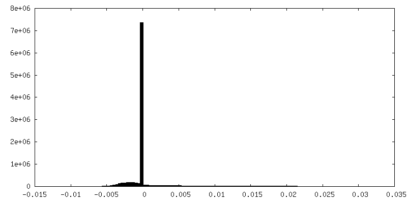
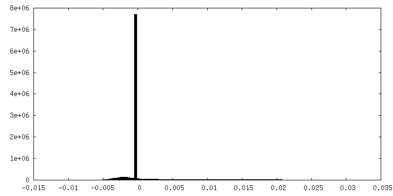
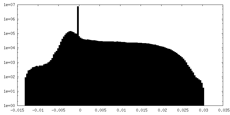
FSC曲線 (解像度の算出)
-
原子モデル構築 1
精密化
空間: REAL / プロトコル: AB INITIO MODEL
得られたモデル
PDB-6r4r: Cryo-EM Structure of the PI3-Kinase SH3 Domain Amyloid Fibril
 ムービー
ムービー コントローラー


 データを開く
 基本情報
基本情報 マップデータ
 試料
試料 キーワード
キーワード 機能・相同性情報
 データ登録者
 ドイツ, 4件
  引用
引用

 構造の表示
構造の表示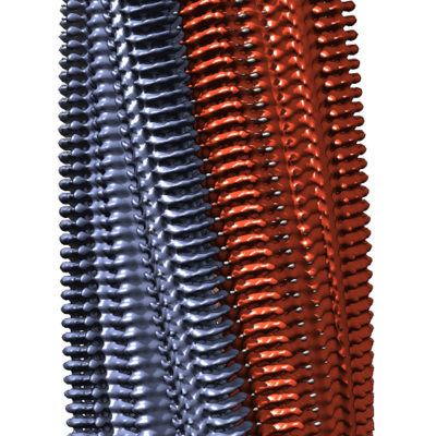
 ダウンロードとリンク
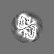
 emd_4727.png
 http://ftp.pdbj.org/pub/emdb/structures/EMD-4727


 Z (Sec.)
 Y (Row.)
 X (Col.)


 試料の構成要素
 解析
解析 電子顕微鏡法
 FIELD EMISSION GUN