 Movie
Movie Controller

[English] 日本語
 Yorodumi
- PDB-4atp: Structure of GABA-transaminase A1R958 from Arthrobacter aurescens... -
+ Open data

- Basic information
| Entry | Database: PDB / ID: 4atp | ||||||
|---|---|---|---|---|---|---|---|
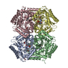
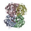
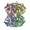
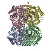
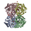
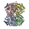
| Title | Structure of GABA-transaminase A1R958 from Arthrobacter aurescens in complex with PLP | ||||||
 Components Components | 4-AMINOBUTYRATE TRANSAMINASE | ||||||
 Keywords Keywords | TRANSFERASE | ||||||
| Function / homology |  Function and homology information Function and homology information5-aminovalerate:2-oxoglutarate transaminase activity / (S)-3-amino-2-methylpropionate:2-oxoglutarate transaminase activity / (S)-3-amino-2-methylpropionate transaminase / 4-aminobutyrate-2-oxoglutarate transaminase / 4-aminobutyrate:2-oxoglutarate transaminase activity / : / pyridoxal phosphate binding / identical protein binding Similarity search - Function | ||||||
| Biological species |  ARTHROBACTER AURESCENS (bacteria) ARTHROBACTER AURESCENS (bacteria) | ||||||
| Method |  X-RAY DIFFRACTION / SYNCHROTRON / MOLECULAR REPLACEMENT / Resolution: 2.8 Å X-RAY DIFFRACTION / SYNCHROTRON / MOLECULAR REPLACEMENT / Resolution: 2.8 Å | ||||||
 Authors Authors | Bruce, H. / Tuan, A.N. / Mangas Sanchez, J. / Hart, S. / Turkenburg, J.P. / Grogan, G. | ||||||
 Citation Citation | Journal: Acta Crystallogr.,Sect.F / Year: 2012 Title: Structures of a Gamma-Aminobutyrate (Gaba) Transaminase from the S-Triazine-Degrading Organism Arthrobacter Aurescens Tc1 in Complex with Plp and with its External Aldimine Plp- Gaba Adduct. Authors: Bruce, H. / Nguyen Tuan, A. / Mangas Sanchez, J. / Leese, C. / Hopwood, J. / Hyde, R. / Hart, S. / Turkenburg, J.P. / Grogan, G. | ||||||
| History |
|
- Structure visualization
| Structure viewer | Molecule: MolmilJmol/JSmol |
|---|
- Downloads & links
-Download
| PDBx/mmCIF format | 4atp.cif.gz | 926.4 KB | Display | PDBx/mmCIF format |
|---|---|---|---|---|
| PDB format | pdb4atp.ent.gz | 771.5 KB | Display | PDB format |
| PDBx/mmJSON format | 4atp.json.gz | Tree view | PDBx/mmJSON format | |
| Others |  Other downloads Other downloads |
-Validation report
| Arichive directory | https://data.pdbj.org/pub/pdb/validation_reports/at/4atpftp://data.pdbj.org/pub/pdb/validation_reports/at/4atp | HTTPS FTP |
|---|
-Related structure data
| Related structure data | 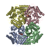 4atqC 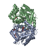 3oksS S: Starting model for refinement C: citing same article ( |
|---|---|
| Similar structure data |
-Links
 PDBj
PDBj- Assembly
| Deposited unit | 
| ||||||||
|---|---|---|---|---|---|---|---|---|---|
| 1 | 
| ||||||||
| 2 | 
| ||||||||
| 3 | 
| ||||||||
| Unit cell |
|
-Components
| #1: Protein | Mass: 47634.102 Da / Num. of mol.: 12 Source method: isolated from a genetically manipulated source Details: COVALENT LINK BETWEEN SIDE CHAN OF LYS295 AND ALDEHYDIC CARBON OF PLP Source: (gene. exp.) ARTHROBACTER AURESCENS (bacteria) / Strain: TC1 / Description: GENOMIC DNA FROM ATCC / Plasmid: PET-YSBLIC-3C / Production host: References: UniProt: A1R958, 4-aminobutyrate-2-oxoglutarate transaminase #2: Chemical | ChemComp-PLP /   Mass: 247.142 Da / Num. of mol.: 12 / Source method: obtained synthetically / Formula: C8H10NO6P Mass: 247.142 Da / Num. of mol.: 12 / Source method: obtained synthetically / Formula: C8H10NO6P#3: Water | ChemComp-HOH / |  Mass: 18.015 Da / Num. of mol.: 746 / Source method: isolated from a natural source / Formula: H2O Mass: 18.015 Da / Num. of mol.: 746 / Source method: isolated from a natural source / Formula: H2ONonpolymer details | PYRIDOXAL-5'-PHOSPHATE (PLP): COVALENTLY | Sequence details | UNIPROT SEQUENCE SUGGESTS POSITION 113 IS ALA BUT DENSITY STRONGLY SUGGESTS THR | |
|---|
-Experimental details
-Experiment
| Experiment | Method: X-RAY DIFFRACTION / Number of used crystals: 1 |
|---|
- Sample preparation
| Crystal | Density Matthews: 2.42 Å3/Da / Density % sol: 49 % / Description: NONE |
|---|---|
| Crystal grow | pH: 7.5 Details: 28% (W/V) PEG 3350, 0.2 M AMMONIUM ACETATE, 3% DIOXANE, 1 MM PLP, 100 MM BIS-TRIS PROPANE PH 7.5, PROTEIN AT 10 MG PER ML. |
-Data collection
| Diffraction | Mean temperature: 120 K |
|---|---|
| Diffraction source | Source: SYNCHROTRON / Site: Diamond  / Beamline: I04 / Wavelength: 0.9795 / Beamline: I04 / Wavelength: 0.9795 |
| Detector | Type: ADSC CCD / Detector: CCD / Date: Feb 12, 2012 |
| Radiation | Protocol: SINGLE WAVELENGTH / Monochromatic (M) / Laue (L): M / Scattering type: x-ray |
| Radiation wavelength | Wavelength: 0.9795 Å / Relative weight: 1 |
| Reflection | Resolution: 2.8→89.84 Å / Num. obs: 125730 / % possible obs: 100 % / Observed criterion σ(I): 2 / Redundancy: 7.4 % / Rmerge(I) obs: 0.18 / Net I/σ(I): 10.5 |
| Reflection shell | Resolution: 2.8→2.87 Å / Redundancy: 7.6 % / Rmerge(I) obs: 0.72 / Mean I/σ(I) obs: 2.6 / % possible all: 100 |
- Processing
| Software |
| ||||||||||||||||||||||||||||||||||||||||||||||||||||||||||||||||||||||||||||||||||||||||||||||||||||||||||||||||||||||||||||||||||||||||||||||||||||||||||||||||||||||||||||||||||||||
|---|---|---|---|---|---|---|---|---|---|---|---|---|---|---|---|---|---|---|---|---|---|---|---|---|---|---|---|---|---|---|---|---|---|---|---|---|---|---|---|---|---|---|---|---|---|---|---|---|---|---|---|---|---|---|---|---|---|---|---|---|---|---|---|---|---|---|---|---|---|---|---|---|---|---|---|---|---|---|---|---|---|---|---|---|---|---|---|---|---|---|---|---|---|---|---|---|---|---|---|---|---|---|---|---|---|---|---|---|---|---|---|---|---|---|---|---|---|---|---|---|---|---|---|---|---|---|---|---|---|---|---|---|---|---|---|---|---|---|---|---|---|---|---|---|---|---|---|---|---|---|---|---|---|---|---|---|---|---|---|---|---|---|---|---|---|---|---|---|---|---|---|---|---|---|---|---|---|---|---|---|---|---|---|
| Refinement | Method to determine structure: MOLECULAR REPLACEMENT Starting model: PDB ENTRY 3OKS Resolution: 2.8→89.84 Å / Cor.coef. Fo:Fc: 0.919 / Cor.coef. Fo:Fc free: 0.889 / SU B: 12.665 / SU ML: 0.25 / Cross valid method: THROUGHOUT / ESU R Free: 0.364 / Stereochemistry target values: MAXIMUM LIKELIHOOD / Details: HYDROGENS HAVE BEEN ADDED IN THE RIDING POSITIONS.
| ||||||||||||||||||||||||||||||||||||||||||||||||||||||||||||||||||||||||||||||||||||||||||||||||||||||||||||||||||||||||||||||||||||||||||||||||||||||||||||||||||||||||||||||||||||||
| Solvent computation | Ion probe radii: 0.8 Å / Shrinkage radii: 0.8 Å / VDW probe radii: 1.2 Å / Solvent model: MASK | ||||||||||||||||||||||||||||||||||||||||||||||||||||||||||||||||||||||||||||||||||||||||||||||||||||||||||||||||||||||||||||||||||||||||||||||||||||||||||||||||||||||||||||||||||||||
| Displacement parameters | Biso mean: 23.221 Å2
| ||||||||||||||||||||||||||||||||||||||||||||||||||||||||||||||||||||||||||||||||||||||||||||||||||||||||||||||||||||||||||||||||||||||||||||||||||||||||||||||||||||||||||||||||||||||
| Refinement step | Cycle: LAST / Resolution: 2.8→89.84 Å
| ||||||||||||||||||||||||||||||||||||||||||||||||||||||||||||||||||||||||||||||||||||||||||||||||||||||||||||||||||||||||||||||||||||||||||||||||||||||||||||||||||||||||||||||||||||||
| Refine LS restraints |
|