 Movie
Movie Controller

[English] 日本語
 Yorodumi
- PDB-1szk: The structure of gamma-aminobutyrate aminotransferase mutant: E211S -
+ Open data

- Basic information
| Entry | Database: PDB / ID: 1szk | ||||||
|---|---|---|---|---|---|---|---|
| Title | The structure of gamma-aminobutyrate aminotransferase mutant: E211S | ||||||
 Components Components | 4-aminobutyrate aminotransferase | ||||||
 Keywords Keywords | TRANSFERASE / GABA-AT | ||||||
| Function / homology |  Function and homology information Function and homology information5-aminovalerate transaminase / 5-aminovalerate:2-oxoglutarate transaminase activity / 4-aminobutyrate-2-oxoglutarate transaminase / 4-aminobutyrate:2-oxoglutarate transaminase activity / N2-acetyl-L-ornithine:2-oxoglutarate 5-transaminase activity / GABA catabolic process / : / pyridoxal phosphate binding / protein homodimerization activity / cytosol Similarity search - Function | ||||||
| Biological species |  | ||||||
| Method |  X-RAY DIFFRACTION / MOLECULAR REPLACEMENT / Resolution: 2.52 Å X-RAY DIFFRACTION / MOLECULAR REPLACEMENT / Resolution: 2.52 Å | ||||||
 Authors Authors | Liu, W. / Peterson, P.E. / Langston, J.A. / Jin, X. / Fisher, A.J. / Toney, M.D. | ||||||
 Citation Citation | Journal: Biochemistry / Year: 2005 Title: Kinetic and Crystallographic Analysis of Active Site Mutants of Escherichia coligamma-Aminobutyrate Aminotransferase. Authors: Liu, W. / Peterson, P.E. / Langston, J.A. / Jin, X. / Zhou, X. / Fisher, A.J. / Toney, M.D. | ||||||
| History |
|
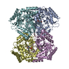
- Structure visualization
| Structure viewer | Molecule: MolmilJmol/JSmol |
|---|
- Downloads & links
-Download
| PDBx/mmCIF format | 1szk.cif.gz | 335.7 KB | Display | PDBx/mmCIF format |
|---|---|---|---|---|
| PDB format | pdb1szk.ent.gz | 276 KB | Display | PDB format |
| PDBx/mmJSON format | 1szk.json.gz | Tree view | PDBx/mmJSON format | |
| Others |  Other downloads Other downloads |
-Validation report
| Arichive directory | https://data.pdbj.org/pub/pdb/validation_reports/sz/1szkftp://data.pdbj.org/pub/pdb/validation_reports/sz/1szk | HTTPS FTP |
|---|
-Related structure data
| Related structure data |  1szsC  1szuC  1sf2S S: Starting model for refinement C: citing same article ( |
|---|---|
| Similar structure data |

-Links
 PDBj
PDBj- Assembly
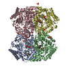
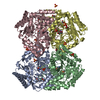
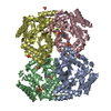
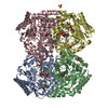
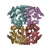
| Deposited unit | 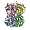
| ||||||||
|---|---|---|---|---|---|---|---|---|---|
| 1 |
| ||||||||
| Unit cell |
|
-Components
| #1: Protein | Mass: 45785.461 Da / Num. of mol.: 4 / Mutation: E211S Source method: isolated from a genetically manipulated source Source: (gene. exp.) References: UniProt: P22256, 4-aminobutyrate-2-oxoglutarate transaminase #2: Chemical | ChemComp-SO4 /   Mass: 96.063 Da / Num. of mol.: 15 / Source method: obtained synthetically / Formula: SO4 Mass: 96.063 Da / Num. of mol.: 15 / Source method: obtained synthetically / Formula: SO4#3: Chemical | ChemComp-EDO /   Mass: 62.068 Da / Num. of mol.: 13 / Source method: obtained synthetically / Formula: C2H6O2 Mass: 62.068 Da / Num. of mol.: 13 / Source method: obtained synthetically / Formula: C2H6O2#4: Chemical | ChemComp-PMP /   Mass: 248.173 Da / Num. of mol.: 4 / Source method: obtained synthetically / Formula: C8H13N2O5P Mass: 248.173 Da / Num. of mol.: 4 / Source method: obtained synthetically / Formula: C8H13N2O5P#5: Water | ChemComp-HOH / |  Mass: 18.015 Da / Num. of mol.: 673 / Source method: isolated from a natural source / Formula: H2O Mass: 18.015 Da / Num. of mol.: 673 / Source method: isolated from a natural source / Formula: H2O |
|---|
-Experimental details
-Experiment
| Experiment | Method: X-RAY DIFFRACTION / Number of used crystals: 1 |
|---|
- Sample preparation
| Crystal | Density Matthews: 2.78 Å3/Da / Density % sol: 55.72 % |
|---|---|
| Crystal grow | Temperature: 293 K / Method: vapor diffusion, hanging drop / pH: 7.6 Details: HEPES, Ammonium Sulfate, PLP, pH 7.6, VAPOR DIFFUSION, HANGING DROP, temperature 293K |
-Data collection
| Diffraction | Mean temperature: 100 K |
|---|---|
| Diffraction source | Source: ROTATING ANODE / Type: RIGAKU RU300 / Wavelength: 1.54 Å |
| Detector | Type: RIGAKU RAXIS IV / Detector: IMAGE PLATE / Date: Aug 10, 2000 |
| Radiation | Monochromator: Ni Filter / Protocol: SINGLE WAVELENGTH / Monochromatic (M) / Laue (L): M / Scattering type: x-ray |
| Radiation wavelength | Wavelength: 1.54 Å / Relative weight: 1 |
| Reflection | Resolution: 2.52→30 Å / Num. all: 71654 / Num. obs: 69873 / % possible obs: 97.5 % / Observed criterion σ(F): 2 / Observed criterion σ(I): 2 / Redundancy: 5.5 % / Rsym value: 0.084 / Net I/σ(I): 12.4 |
| Reflection shell | Resolution: 2.52→2.59 Å / % possible all: 97.5 |
- Processing
| Software |
| ||||||||||||||||||||||||||||
|---|---|---|---|---|---|---|---|---|---|---|---|---|---|---|---|---|---|---|---|---|---|---|---|---|---|---|---|---|---|
| Refinement | Method to determine structure: MOLECULAR REPLACEMENT Starting model: pdb entry 1SF2 Resolution: 2.52→30 Å / σ(F): 2 / Stereochemistry target values: Engh & Huber
| ||||||||||||||||||||||||||||
| Displacement parameters |
| ||||||||||||||||||||||||||||
| Refinement step | Cycle: LAST / Resolution: 2.52→30 Å
| ||||||||||||||||||||||||||||
| Refine LS restraints |
| ||||||||||||||||||||||||||||
| Xplor file |
|
