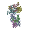
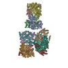
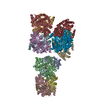
Journal: Nat Commun / Year: 2019 Title: Atomic structure of PI3-kinase SH3 amyloid fibrils by cryo-electron microscopy. Authors: Christine Röder / Nicola Vettore / Lena N Mangels / Lothar Gremer / Raimond B G Ravelli / Dieter Willbold / Wolfgang Hoyer / Alexander K Buell / Gunnar F Schröder / Abstract: High resolution structural information on amyloid fibrils is crucial for the understanding of their formation mechanisms and for the rational design of amyloid inhibitors in the context of protein ...High resolution structural information on amyloid fibrils is crucial for the understanding of their formation mechanisms and for the rational design of amyloid inhibitors in the context of protein misfolding diseases. The Src-homology 3 domain of phosphatidyl-inositol-3-kinase (PI3K-SH3) is a model amyloid system that plays a pivotal role in our basic understanding of protein misfolding and aggregation. Here, we present the atomic model of the PI3K-SH3 amyloid fibril with a resolution determined to 3.4 Å by cryo-electron microscopy (cryo-EM). The fibril is composed of two intertwined protofilaments that create an interface spanning 13 residues from each monomer. The model comprises residues 1-77 out of 86 amino acids in total, with the missing residues located in the highly flexible C-terminus. The fibril structure allows us to rationalise the effects of chemically conservative point mutations as well as of the previously reported sequence perturbations on PI3K-SH3 fibril formation and growth.
History
Deposition
Mar 23, 2019
-
Header (metadata) release
Aug 28, 2019
-
Map release
Aug 28, 2019
-
Update
May 15, 2024
-
Current status
May 15, 2024
Processing site: PDBe / Status: Released
-
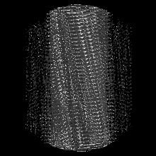
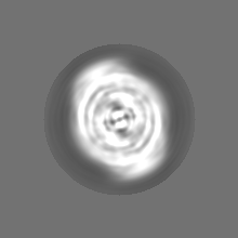
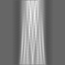
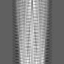
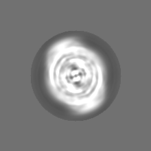
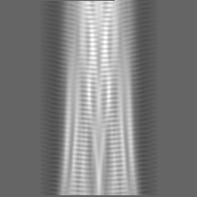
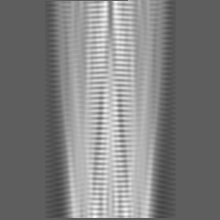
Structure visualization
Movie
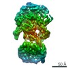
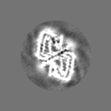
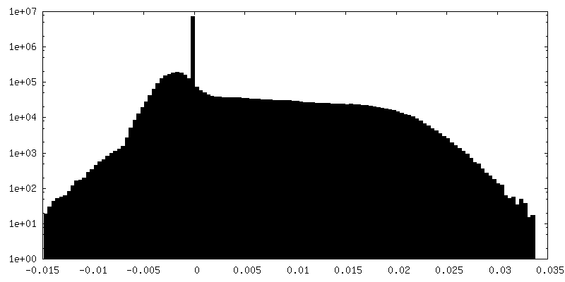
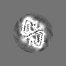
Surface view with section colored by density value
Model: Quantifoil R1.2/1.3 / Material: COPPER / Mesh: 300 / Support film - Material: CARBON / Support film - topology: HOLEY / Pretreatment - Type: GLOW DISCHARGE
Vitrification
Cryogen name: ETHANE
-
Electron microscopy
Microscope
FEI TECNAI ARCTICA
Image recording
Film or detector model: FEI FALCON III (4k x 4k) / Detector mode: COUNTING / Digitization - Dimensions - Width: 4096 pixel / Digitization - Dimensions - Height: 4096 pixel / Number real images: 622 / Average exposure time: 65.0 sec. / Average electron dose: 26.0 e/Å2
Electron beam
Acceleration voltage: 200 kV / Electron source: FIELD EMISSION GUN
Electron optics
Illumination mode: FLOOD BEAM / Imaging mode: BRIGHT FIELD / Cs: 2.7 mm
Experimental equipment
Model: Talos Arctica / Image courtesy: FEI Company
+
Image processing
Final reconstruction
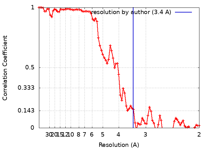
Applied symmetry - Helical parameters - Δz: 2.3548 Å Applied symmetry - Helical parameters - Δ&Phi: 179.436 ° Applied symmetry - Helical parameters - Axial symmetry: C1 (asymmetric) Resolution.type: BY AUTHOR / Resolution: 3.4 Å / Resolution method: FSC 0.143 CUT-OFF / Software - Name: RELION (ver. 2) Details: For the even/odd test, the segment images were split by entire fibrils and refined separately for 25 iterations using the same reference density (low-passed filtered to 20 A). Number images used: 27681
Segment selection
Number selected: 103733 / Details: Filaments were picked manually in Relion2
Startup model
Type of model: NONE
Final angle assignment
Type: NOT APPLICABLE / Software - Name: RELION (ver. 2)
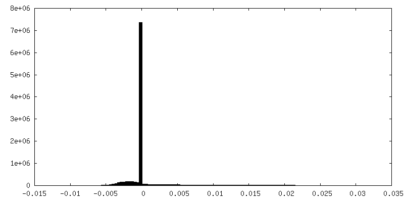
FSC plot (resolution estimation)
-
Atomic model buiding 1
Refinement
Space: REAL / Protocol: AB INITIO MODEL
Output model
PDB-6r4r: Cryo-EM Structure of the PI3-Kinase SH3 Domain Amyloid Fibril
+
About Yorodumi
-
News
-
Feb 9, 2022. New format data for meta-information of EMDB entries
New format data for meta-information of EMDB entries
Version 3 of the EMDB header file is now the official format.
The previous official version 1.9 will be removed from the archive.
In the structure databanks used in Yorodumi, some data are registered as the other names, "COVID-19 virus" and "2019-nCoV". Here are the details of the virus and the list of structure data.
Jan 31, 2019. EMDB accession codes are about to change! (news from PDBe EMDB page)
EMDB accession codes are about to change! (news from PDBe EMDB page)
The allocation of 4 digits for EMDB accession codes will soon come to an end. Whilst these codes will remain in use, new EMDB accession codes will include an additional digit and will expand incrementally as the available range of codes is exhausted. The current 4-digit format prefixed with “EMD-” (i.e. EMD-XXXX) will advance to a 5-digit format (i.e. EMD-XXXXX), and so on. It is currently estimated that the 4-digit codes will be depleted around Spring 2019, at which point the 5-digit format will come into force.
The EM Navigator/Yorodumi systems omit the EMD- prefix.
Related info.:Q: What is EMD? / ID/Accession-code notation in Yorodumi/EM Navigator
Yorodumi is a browser for structure data from EMDB, PDB, SASBDB, etc.
This page is also the successor to EM Navigator detail page, and also detail information page/front-end page for Omokage search.
The word "yorodu" (or yorozu) is an old Japanese word meaning "ten thousand". "mi" (miru) is to see.
Related info.:EMDB / PDB / SASBDB / Comparison of 3 databanks / Yorodumi Search / Aug 31, 2016. New EM Navigator & Yorodumi / Yorodumi Papers / Jmol/JSmol / Function and homology information / Changes in new EM Navigator and Yorodumi
 Movie
Movie Controller


 Open data
 Basic information
 Map data
 Sample
 Keywords
 Function and homology information
 Authors
 Germany, 4 items
  Citation

 Structure visualization
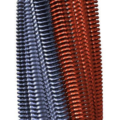
 Downloads & links
 emd_4727.png
 http://ftp.pdbj.org/pub/emdb/structures/EMD-4727


 Z (Sec.)
 Y (Row.)
 X (Col.)


 Sample components
 Processing
 Electron microscopy
 FIELD EMISSION GUN