+ Open data
- Basic information
| Entry | Database: EMDB / ID: EMD-4520 | |||||||||
|---|---|---|---|---|---|---|---|---|---|---|
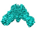
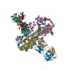
| Title | EM structure of a EBOV-GP bound to 3T0331 neutralizing antibody | |||||||||
 Map data Map data | Local filtered map | |||||||||
 Sample Sample |
| |||||||||
 Keywords Keywords | Antibody / immune response / viral infection / EBOV / VIRAL PROTEIN | |||||||||
| Function / homology |  Function and homology information Function and homology informationsymbiont-mediated killing of host cell / host cell endoplasmic reticulum / viral budding from plasma membrane / clathrin-dependent endocytosis of virus by host cell / symbiont-mediated-mediated suppression of host tetherin activity / entry receptor-mediated virion attachment to host cell / host cell cytoplasm / symbiont-mediated suppression of host innate immune response / membrane raft / fusion of virus membrane with host endosome membrane ...symbiont-mediated killing of host cell / host cell endoplasmic reticulum / viral budding from plasma membrane / clathrin-dependent endocytosis of virus by host cell / symbiont-mediated-mediated suppression of host tetherin activity / entry receptor-mediated virion attachment to host cell / host cell cytoplasm / symbiont-mediated suppression of host innate immune response / membrane raft / fusion of virus membrane with host endosome membrane / viral envelope / symbiont entry into host cell / lipid binding / host cell plasma membrane / virion membrane / extracellular region / identical protein binding Similarity search - Function | |||||||||
| Biological species |   Homo sapiens (human) Homo sapiens (human) | |||||||||
| Method | single particle reconstruction / cryo EM / Resolution: 3.1 Å | |||||||||
 Authors Authors | Diskin R / Cohen-Dvashi H | |||||||||
 Citation Citation |  Journal: Nat Med / Year: 2019 Journal: Nat Med / Year: 2019Title: Polyclonal and convergent antibody response to Ebola virus vaccine rVSV-ZEBOV. Authors: Stefanie A Ehrhardt / Matthias Zehner / Verena Krähling / Hadas Cohen-Dvashi / Christoph Kreer / Nadav Elad / Henning Gruell / Meryem S Ercanoglu / Philipp Schommers / Lutz Gieselmann / ...Authors: Stefanie A Ehrhardt / Matthias Zehner / Verena Krähling / Hadas Cohen-Dvashi / Christoph Kreer / Nadav Elad / Henning Gruell / Meryem S Ercanoglu / Philipp Schommers / Lutz Gieselmann / Ralf Eggeling / Christine Dahlke / Timo Wolf / Nico Pfeifer / Marylyn M Addo / Ron Diskin / Stephan Becker / Florian Klein /   Abstract: Recombinant vesicular stomatitis virus-Zaire Ebola virus (rVSV-ZEBOV) is the most advanced Ebola virus vaccine candidate and is currently being used to combat the outbreak of Ebola virus disease (EVD) ...Recombinant vesicular stomatitis virus-Zaire Ebola virus (rVSV-ZEBOV) is the most advanced Ebola virus vaccine candidate and is currently being used to combat the outbreak of Ebola virus disease (EVD) in the Democratic Republic of the Congo (DRC). Here we examine the humoral immune response in a subset of human volunteers enrolled in a phase 1 rVSV-ZEBOV vaccination trial by performing comprehensive single B cell and electron microscopy structure analyses. Four studied vaccinees show polyclonal, yet reproducible and convergent B cell responses with shared sequence characteristics. EBOV-targeting antibodies cross-react with other Ebolavirus species, and detailed epitope mapping revealed overlapping target epitopes with antibodies isolated from EVD survivors. Moreover, in all vaccinees, we detected highly potent EBOV-neutralizing antibodies with activities comparable or superior to the monoclonal antibodies currently used in clinical trials. These include antibodies combining the IGHV3-15/IGLV1-40 immunoglobulin gene segments that were identified in all investigated individuals. Our findings will help to evaluate and direct current and future vaccination strategies and offer opportunities for novel EVD therapies. | |||||||||
| History |
|
- Structure visualization
| Movie |
 Movie viewer Movie viewer |
|---|---|
| Structure viewer | EM map:  SurfView SurfView Molmil Molmil Jmol/JSmol Jmol/JSmol |
| Supplemental images |
- Downloads & links
-EMDB archive
| Map data |  emd_4520.map.gz emd_4520.map.gz | 5.9 MB |  EMDB map data format EMDB map data format | |
|---|---|---|---|---|
| Header (meta data) |  emd-4520-v30.xml emd-4520-v30.xml emd-4520.xml emd-4520.xml | 21 KB 21 KB | Display Display |  EMDB header EMDB header |
| FSC (resolution estimation) |  emd_4520_fsc.xml emd_4520_fsc.xml | 15 KB | Display |  FSC data file FSC data file |
| Images |  emd_4520.png emd_4520.png | 174 KB | ||
| Filedesc metadata |  emd-4520.cif.gz emd-4520.cif.gz | 6.7 KB | ||
| Others |  emd_4520_half_map_1.map.gz emd_4520_half_map_1.map.gz emd_4520_half_map_2.map.gz emd_4520_half_map_2.map.gz | 165.4 MB 165.4 MB | ||
| Archive directory |  http://ftp.pdbj.org/pub/emdb/structures/EMD-4520 http://ftp.pdbj.org/pub/emdb/structures/EMD-4520 ftp://ftp.pdbj.org/pub/emdb/structures/EMD-4520 ftp://ftp.pdbj.org/pub/emdb/structures/EMD-4520 | HTTPS FTP |
-Validation report
| Summary document |  emd_4520_validation.pdf.gz emd_4520_validation.pdf.gz | 899.1 KB | Display |  EMDB validaton report EMDB validaton report |
|---|---|---|---|---|
| Full document |  emd_4520_full_validation.pdf.gz emd_4520_full_validation.pdf.gz | 898.7 KB | Display | |
| Data in XML |  emd_4520_validation.xml.gz emd_4520_validation.xml.gz | 20.2 KB | Display | |
| Data in CIF |  emd_4520_validation.cif.gz emd_4520_validation.cif.gz | 26.9 KB | Display | |
| Arichive directory |  https://ftp.pdbj.org/pub/emdb/validation_reports/EMD-4520 https://ftp.pdbj.org/pub/emdb/validation_reports/EMD-4520 ftp://ftp.pdbj.org/pub/emdb/validation_reports/EMD-4520 ftp://ftp.pdbj.org/pub/emdb/validation_reports/EMD-4520 | HTTPS FTP |
-Related structure data
| Related structure data |  6qd7MC  4521C  6qd8C M: atomic model generated by this map C: citing same article ( |
|---|---|
| Similar structure data |
- Links
Links
| EMDB pages |  EMDB (EBI/PDBe) / EMDB (EBI/PDBe) /  EMDataResource EMDataResource |
|---|---|
| Related items in Molecule of the Month |
- Map
Map
| File |  Download / File: emd_4520.map.gz / Format: CCP4 / Size: 178 MB / Type: IMAGE STORED AS FLOATING POINT NUMBER (4 BYTES) Download / File: emd_4520.map.gz / Format: CCP4 / Size: 178 MB / Type: IMAGE STORED AS FLOATING POINT NUMBER (4 BYTES) | ||||||||||||||||||||||||||||||||||||||||||||||||||||||||||||||||||||
|---|---|---|---|---|---|---|---|---|---|---|---|---|---|---|---|---|---|---|---|---|---|---|---|---|---|---|---|---|---|---|---|---|---|---|---|---|---|---|---|---|---|---|---|---|---|---|---|---|---|---|---|---|---|---|---|---|---|---|---|---|---|---|---|---|---|---|---|---|---|
| Annotation | Local filtered map | ||||||||||||||||||||||||||||||||||||||||||||||||||||||||||||||||||||
| Projections & slices | Image control
Images are generated by Spider. | ||||||||||||||||||||||||||||||||||||||||||||||||||||||||||||||||||||
| Voxel size | X=Y=Z: 0.85 Å | ||||||||||||||||||||||||||||||||||||||||||||||||||||||||||||||||||||
| Density |
| ||||||||||||||||||||||||||||||||||||||||||||||||||||||||||||||||||||
| Symmetry | Space group: 1 | ||||||||||||||||||||||||||||||||||||||||||||||||||||||||||||||||||||
| Details | EMDB XML:
CCP4 map header:
| ||||||||||||||||||||||||||||||||||||||||||||||||||||||||||||||||||||
-Supplemental data
-Half map: Half map A
| File | emd_4520_half_map_1.map | ||||||||||||
|---|---|---|---|---|---|---|---|---|---|---|---|---|---|
| Annotation | Half map A | ||||||||||||
| Projections & Slices |
| ||||||||||||
| Density Histograms |
-Half map: Half map B
| File | emd_4520_half_map_2.map | ||||||||||||
|---|---|---|---|---|---|---|---|---|---|---|---|---|---|
| Annotation | Half map B | ||||||||||||
| Projections & Slices |
| ||||||||||||
| Density Histograms |
- Sample components
-Entire : EBOV-GP in complex with neutralizing 3T0331 antibody
| Entire | Name: EBOV-GP in complex with neutralizing 3T0331 antibody |
|---|---|
| Components |
|
-Supramolecule #1: EBOV-GP in complex with neutralizing 3T0331 antibody
| Supramolecule | Name: EBOV-GP in complex with neutralizing 3T0331 antibody / type: complex / ID: 1 / Parent: 0 / Macromolecule list: #1-#4 |
|---|
-Supramolecule #2: EBOV-GP
| Supramolecule | Name: EBOV-GP / type: complex / ID: 2 / Parent: 1 / Macromolecule list: #3-#4 |
|---|---|
| Source (natural) | Organism:  |
-Supramolecule #3: neutralizing 3T0331 antibody
| Supramolecule | Name: neutralizing 3T0331 antibody / type: complex / ID: 3 / Parent: 1 / Macromolecule list: #1-#2 |
|---|---|
| Source (natural) | Organism:  Homo sapiens (human) Homo sapiens (human) |
-Macromolecule #1: Light chain
| Macromolecule | Name: Light chain / type: protein_or_peptide / ID: 1 / Number of copies: 3 / Enantiomer: LEVO |
|---|---|
| Source (natural) | Organism:  Homo sapiens (human) Homo sapiens (human) |
| Molecular weight | Theoretical: 23.310885 KDa |
| Recombinant expression | Organism:  Homo sapiens (human) Homo sapiens (human) |
| Sequence | String: EIVLTQSPAT LSLSPGERAT LSCRASQSVS TYLAWYQHQP GQAPRLLIYE ASNRATGIPA RFSGSGSGTE FTLTISSLEP EDVAVYYCQ QRASWPLTFG GGTKVEIKRT VAAPSVFIFP PSDEQLKSGT ASVVCLLNNF YPREAKVQWK VDNALQSGNS Q ESVTEQDS ...String: EIVLTQSPAT LSLSPGERAT LSCRASQSVS TYLAWYQHQP GQAPRLLIYE ASNRATGIPA RFSGSGSGTE FTLTISSLEP EDVAVYYCQ QRASWPLTFG GGTKVEIKRT VAAPSVFIFP PSDEQLKSGT ASVVCLLNNF YPREAKVQWK VDNALQSGNS Q ESVTEQDS KDSTYSLSST LTLSKADYEK HKVYACEVTH QGLSSPVTKS FNRGEC |
-Macromolecule #2: Heavy chain
| Macromolecule | Name: Heavy chain / type: protein_or_peptide / ID: 2 / Number of copies: 3 / Enantiomer: LEVO |
|---|---|
| Source (natural) | Organism:  Homo sapiens (human) Homo sapiens (human) |
| Molecular weight | Theoretical: 23.673553 KDa |
| Recombinant expression | Organism:  Homo sapiens (human) Homo sapiens (human) |
| Sequence | String: EVQLLESGGG LVQPGGSLRL SCEASGFPLR DYAMSWVRQA PGRGLQWVST IGGNDNAANY ADSVKGRFTV SRDNSKSTIY LQMNSLRAE DTALYFCAKS VRLSRPSPFD LWGQGSLVTV SSASTKGPSV FPLAPSSKST SGGTAALGCL VKDYFPEPVT V SWNSGALT ...String: EVQLLESGGG LVQPGGSLRL SCEASGFPLR DYAMSWVRQA PGRGLQWVST IGGNDNAANY ADSVKGRFTV SRDNSKSTIY LQMNSLRAE DTALYFCAKS VRLSRPSPFD LWGQGSLVTV SSASTKGPSV FPLAPSSKST SGGTAALGCL VKDYFPEPVT V SWNSGALT SGVHTFPAVL QSSGLYSLSS VVTVPSSSLG TQTYICNVNH KPSNTKVDKR VEPKSC |
-Macromolecule #3: Envelope glycoprotein,Virion spike glycoprotein,EBOV-GP1
| Macromolecule | Name: Envelope glycoprotein,Virion spike glycoprotein,EBOV-GP1 type: protein_or_peptide / ID: 3 / Number of copies: 3 / Enantiomer: LEVO |
|---|---|
| Source (natural) | Organism:  |
| Molecular weight | Theoretical: 34.887578 KDa |
| Recombinant expression | Organism:  Homo sapiens (human) Homo sapiens (human) |
| Sequence | String: GSRSIPLGVI HNSALQVSDV DKLVCRDKLS STNQLRSVGL NLEGNGVATD VPSATKRWGF RSGVPPKVVN YEAGEWAENC YNLEIKKPD GSECLPAAPD GIRGFPRCRY VHKVSGTGPC AGDFAFHKEG AFFLYDRLAS TVIYRGTTFA EGVVAFLILP Q AKKDFFSS ...String: GSRSIPLGVI HNSALQVSDV DKLVCRDKLS STNQLRSVGL NLEGNGVATD VPSATKRWGF RSGVPPKVVN YEAGEWAENC YNLEIKKPD GSECLPAAPD GIRGFPRCRY VHKVSGTGPC AGDFAFHKEG AFFLYDRLAS TVIYRGTTFA EGVVAFLILP Q AKKDFFSS HPLREPVNAT EDPSSGYYST TIRYQATGFG TNETEYLFEV DNLTYVQLES RFTPQFLLQL NETIYTSGKR SN TTGKLIW KVNPEIDTTI GEWAFWETKK NLTRKIRSEE LSFTVV(UNK)(UNK)(UNK)(UNK) (UNK)(UNK)(UNK) (UNK)(UNK)(UNK)(UNK)(UNK)(UNK) (UNK)(UNK)(UNK)(UNK)(UNK)(UNK)(UNK)(UNK)(UNK)(UNK) (UNK)(UNK)(UNK)(UNK)(UNK)(UNK)(UNK)(UNK)(UNK) (UNK)(UNK)(UNK)(UNK)(UNK)(UNK)(UNK) UniProtKB: Envelope glycoprotein |
-Macromolecule #4: Envelope glycoprotein
| Macromolecule | Name: Envelope glycoprotein / type: protein_or_peptide / ID: 4 / Number of copies: 3 / Enantiomer: LEVO |
|---|---|
| Source (natural) | Organism:  |
| Molecular weight | Theoretical: 18.989391 KDa |
| Recombinant expression | Organism:  Homo sapiens (human) Homo sapiens (human) |
| Sequence | String: EAIVNAQPKC NPNLHYWTTQ DEGAAIGLAW IPYFGPAAEG IYIEGLMHNQ DGLICGLRQL ANETTQALQL FLRATTELRT FSILNRKAI DFLLQRWGGT CHILGPDCCI EPHDWTKNIT DKIDQIIHDF VDGSGYIPEA PRDGQAYVRK DGEWVLLSTF L GTHHHHHH UniProtKB: Envelope glycoprotein |
-Macromolecule #6: alpha-D-mannopyranose
| Macromolecule | Name: alpha-D-mannopyranose / type: ligand / ID: 6 / Number of copies: 3 / Formula: MAN |
|---|---|
| Molecular weight | Theoretical: 180.156 Da |
| Chemical component information |  ChemComp-MAN: |
-Macromolecule #7: 2-acetamido-2-deoxy-beta-D-glucopyranose
| Macromolecule | Name: 2-acetamido-2-deoxy-beta-D-glucopyranose / type: ligand / ID: 7 / Number of copies: 3 / Formula: NAG |
|---|---|
| Molecular weight | Theoretical: 221.208 Da |
| Chemical component information |  ChemComp-NAG: |
-Experimental details
-Structure determination
| Method | cryo EM |
|---|---|
 Processing Processing | single particle reconstruction |
| Aggregation state | particle |
- Sample preparation
| Buffer | pH: 7.6 |
|---|---|
| Vitrification | Cryogen name: ETHANE |
- Electron microscopy
| Microscope | FEI TITAN KRIOS |
|---|---|
| Image recording | Film or detector model: FEI FALCON III (4k x 4k) / Detector mode: SUPER-RESOLUTION / Average exposure time: 27.0 sec. / Average electron dose: 40.0 e/Å2 |
| Electron beam | Acceleration voltage: 300 kV / Electron source:  FIELD EMISSION GUN FIELD EMISSION GUN |
| Electron optics | Illumination mode: FLOOD BEAM / Imaging mode: BRIGHT FIELD / Cs: 2.7 mm / Nominal magnification: 96000 |
| Sample stage | Specimen holder model: FEI TITAN KRIOS AUTOGRID HOLDER / Cooling holder cryogen: NITROGEN |
| Experimental equipment |  Model: Titan Krios / Image courtesy: FEI Company |
 Movie
Movie Controller


 Z (Sec.)
 Y (Row.)
 X (Col.)


