- EMDB-4164: Cryo-EM structure of the DNA bound DDK phosphorylated MCM double ... -
+
Open data
ID or keywords:
Loading...
-
Basic information
Entry
Database: EMDB / ID: EMD-4164
Title
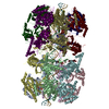
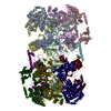
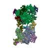
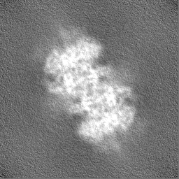
Cryo-EM structure of the DNA bound DDK phosphorylated MCM double hexamer
Map data
None
Sample
Complex: S. cerevisiae DNA bound DDK phosphorylated MCM double hexamer
Function / homology
Function and homology information
MCM core complex / Assembly of the pre-replicative complex / Switching of origins to a post-replicative state / MCM complex binding / nuclear DNA replication / premeiotic DNA replication / pre-replicative complex assembly involved in nuclear cell cycle DNA replication / mitotic DNA replication / Activation of the pre-replicative complex / CMG complex ...MCM core complex / Assembly of the pre-replicative complex / Switching of origins to a post-replicative state / MCM complex binding / nuclear DNA replication / premeiotic DNA replication / pre-replicative complex assembly involved in nuclear cell cycle DNA replication / mitotic DNA replication / Activation of the pre-replicative complex / CMG complex / nuclear pre-replicative complex / MCM complex / Activation of ATR in response to replication stress / DNA replication preinitiation complex / replication fork protection complex / double-strand break repair via break-induced replication / mitotic DNA replication initiation / single-stranded DNA helicase activity / silent mating-type cassette heterochromatin formation / regulation of DNA-templated DNA replication initiation / DNA strand elongation involved in DNA replication / DNA unwinding involved in DNA replication / nuclear replication fork / DNA replication origin binding / DNA replication initiation / subtelomeric heterochromatin formation / DNA helicase activity / helicase activity / transcription elongation by RNA polymerase II / heterochromatin formation / single-stranded DNA binding / chromosome, telomeric region / DNA helicase / chromatin binding / DNA damage response / ATP hydrolysis activity / nucleoplasm / ATP binding / nucleus / metal ion binding / cytoplasm Similarity search - Function
MCM4, winged helix domain / DNA replication licensing factor Mcm5 / DNA replication licensing factor Mcm3 / Mini-chromosome maintenance complex protein 4 / DNA replication licensing factor Mcm6 / DNA replication licensing factor Mcm7 / Mcm6, C-terminal winged-helix domain / MCM6 C-terminal winged-helix domain / DNA replication licensing factor Mcm2 / Mini-chromosome maintenance protein 2 ...MCM4, winged helix domain / DNA replication licensing factor Mcm5 / DNA replication licensing factor Mcm3 / Mini-chromosome maintenance complex protein 4 / DNA replication licensing factor Mcm6 / DNA replication licensing factor Mcm7 / Mcm6, C-terminal winged-helix domain / MCM6 C-terminal winged-helix domain / DNA replication licensing factor Mcm2 / Mini-chromosome maintenance protein 2 / Mini-chromosome maintenance, conserved site / MCM family signature. / MCM N-terminal domain / MCM N-terminal domain / MCM OB domain / MCM OB domain / Mini-chromosome maintenance protein / MCM, AAA-lid domain / MCM P-loop domain / MCM AAA-lid domain / MCM family domain profile. / minichromosome maintenance proteins / MCM domain / Winged helix-like DNA-binding domain superfamily / ATPases associated with a variety of cellular activities / AAA+ ATPase domain / Nucleic acid-binding, OB-fold / P-loop containing nucleoside triphosphate hydrolase Similarity search - Domain/homology
DNA replication licensing factor MCM3 / DNA replication licensing factor MCM2 / Minichromosome maintenance protein 5 / DNA replication licensing factor MCM4 / DNA replication licensing factor MCM7 / DNA replication licensing factor MCM6 Similarity search - Component
Biological species
Saccharomyces cerevisiae (brewer's yeast)
Method
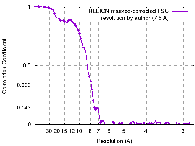
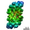
single particle reconstruction / cryo EM / Resolution: 7.5 Å
Journal: Nat Commun / Year: 2017 Title: Cryo-EM structure of a licensed DNA replication origin. Authors: Ferdos Abid Ali / Max E Douglas / Julia Locke / Valerie E Pye / Andrea Nans / John F X Diffley / Alessandro Costa / Abstract: Eukaryotic origins of replication are licensed upon loading of the MCM helicase motor onto DNA. ATP hydrolysis by MCM is required for loading and the post-catalytic MCM is an inactive double hexamer ...Eukaryotic origins of replication are licensed upon loading of the MCM helicase motor onto DNA. ATP hydrolysis by MCM is required for loading and the post-catalytic MCM is an inactive double hexamer that encircles duplex DNA. Origin firing depends on MCM engagement of Cdc45 and GINS to form the CMG holo-helicase. CMG assembly requires several steps including MCM phosphorylation by DDK. To understand origin activation, here we have determined the cryo-EM structures of DNA-bound MCM, either unmodified or phosphorylated, and visualize a phospho-dependent MCM element likely important for Cdc45 recruitment. MCM pore loops touch both the Watson and Crick strands, constraining duplex DNA in a bent configuration. By comparing our new MCM-DNA structure with the structure of CMG-DNA, we suggest how the conformational transition from the loaded, post-catalytic MCM to CMG might promote DNA untwisting and melting at the onset of replication.
History
Deposition
Nov 20, 2017
-
Header (metadata) release
Dec 6, 2017
-
Map release
Dec 6, 2017
-
Update
Jan 24, 2018
-
Current status
Jan 24, 2018
Processing site: PDBe / Status: Released
-
Structure visualization
Movie
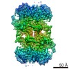
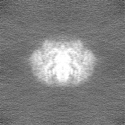
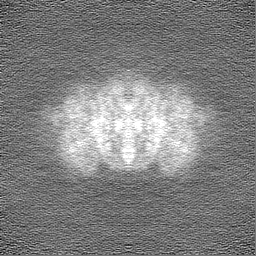
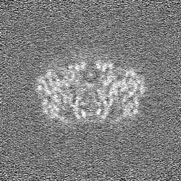
Surface view with section colored by density value
In the structure databanks used in Yorodumi, some data are registered as the other names, "COVID-19 virus" and "2019-nCoV". Here are the details of the virus and the list of structure data.
Jan 31, 2019. EMDB accession codes are about to change! (news from PDBe EMDB page)
EMDB accession codes are about to change! (news from PDBe EMDB page)
The allocation of 4 digits for EMDB accession codes will soon come to an end. Whilst these codes will remain in use, new EMDB accession codes will include an additional digit and will expand incrementally as the available range of codes is exhausted. The current 4-digit format prefixed with “EMD-” (i.e. EMD-XXXX) will advance to a 5-digit format (i.e. EMD-XXXXX), and so on. It is currently estimated that the 4-digit codes will be depleted around Spring 2019, at which point the 5-digit format will come into force.
The EM Navigator/Yorodumi systems omit the EMD- prefix.
Related info.:Q: What is EMD? / ID/Accession-code notation in Yorodumi/EM Navigator
Yorodumi is a browser for structure data from EMDB, PDB, SASBDB, etc.
This page is also the successor to EM Navigator detail page, and also detail information page/front-end page for Omokage search.
The word "yorodu" (or yorozu) is an old Japanese word meaning "ten thousand". "mi" (miru) is to see.
Related info.:EMDB / PDB / SASBDB / Comparison of 3 databanks / Yorodumi Search / Aug 31, 2016. New EM Navigator & Yorodumi / Yorodumi Papers / Jmol/JSmol / Function and homology information / Changes in new EM Navigator and Yorodumi
 Movie
Movie Controller

 Yorodumi
 Open data
 Basic information
 Map data
 Sample
 Function and homology information
 Authors
 Citation
 Structure visualization
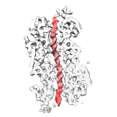
 Downloads & links
 emd_4164.png
 http://ftp.pdbj.org/pub/emdb/structures/EMD-4164
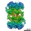


 Z
Z Y
Y X
X


 Sample components
 Processing
 Electron microscopy
 FIELD EMISSION GUN