 Movie
Movie Controller

[English] 日本語
 Yorodumi
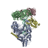
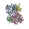
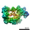
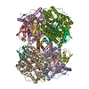
- EMDB-4141: Cryo-EM structure of the E. coli replicative DNA polymerase-clamp... -
+ Open data

- Basic information
| Entry | Database: EMDB / ID: EMD-4141 | |||||||||
|---|---|---|---|---|---|---|---|---|---|---|
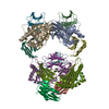
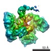
| Title | Cryo-EM structure of the E. coli replicative DNA polymerase-clamp-exonuclase-theta complex bound to DNA in the editing mode | |||||||||
 Map data Map data | Main map | |||||||||
 Sample Sample |
| |||||||||
 Keywords Keywords | DNA editing Proofreading Exonuclease Polymerase / DNA Binding protein | |||||||||
| Function / homology |  Function and homology information Function and homology informationDNA polymerase III, core complex / Hda-beta clamp complex / bacterial-type DNA replication / replication inhibiting complex / DNA polymerase III complex / DNA replication proofreading / lagging strand elongation / replisome / regulation of DNA-templated DNA replication initiation / exonuclease activity ...DNA polymerase III, core complex / Hda-beta clamp complex / bacterial-type DNA replication / replication inhibiting complex / DNA polymerase III complex / DNA replication proofreading / lagging strand elongation / replisome / regulation of DNA-templated DNA replication initiation / exonuclease activity / DNA strand elongation involved in DNA replication / leading strand elongation / error-prone translesion synthesis / 3'-5' exonuclease activity / negative regulation of DNA-templated DNA replication initiation / DNA-templated DNA replication / DNA-directed DNA polymerase / DNA-directed DNA polymerase activity / DNA damage response / protein homodimerization activity / DNA binding / metal ion binding / identical protein binding / cytoplasm / cytosol Similarity search - Function | |||||||||
| Biological species |  | |||||||||
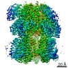
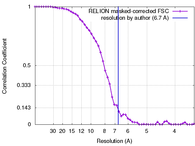
| Method | single particle reconstruction / cryo EM / Resolution: 6.7 Å | |||||||||
 Authors Authors | Fernandez-Leiro R / Conrad J | |||||||||
 Citation Citation | Journal: Nat Struct Mol Biol / Year: 2017 Title: Self-correcting mismatches during high-fidelity DNA replication. Authors: Rafael Fernandez-Leiro / Julian Conrad / Ji-Chun Yang / Stefan M V Freund / Sjors H W Scheres / Meindert H Lamers /  Abstract: Faithful DNA replication is essential to all forms of life and depends on the action of 3'-5' exonucleases that remove misincorporated nucleotides from the newly synthesized strand. However, how the ...Faithful DNA replication is essential to all forms of life and depends on the action of 3'-5' exonucleases that remove misincorporated nucleotides from the newly synthesized strand. However, how the DNA is transferred from the polymerase to the exonuclease active site is not known. Here we present the cryo-EM structure of the editing mode of the catalytic core of the Escherichia coli replisome, revealing a dramatic distortion of the DNA whereby the polymerase thumb domain acts as a wedge that separates the two DNA strands. Importantly, NMR analysis of the DNA substrate shows that the presence of a mismatch increases the fraying of the DNA, thus enabling it to reach the exonuclease active site. Therefore the mismatch corrects itself, whereas the exonuclease subunit plays a passive role. Hence, our work provides unique insights into high-fidelity replication and establishes a new paradigm for the correction of misincorporated nucleotides. | |||||||||
| History |
|
- Structure visualization
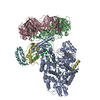
| Movie |
Movie viewer |
|---|---|
| Structure viewer | EM map: SurfViewMolmilJmol/JSmol |
| Supplemental images |

- Downloads & links
-EMDB archive
| Map data | emd_4141.map.gz | 9.7 MB | EMDB map data format | |
|---|---|---|---|---|
| Header (meta data) | emd-4141-v30.xmlemd-4141.xml | 27.4 KB 27.4 KB | Display Display | EMDB header |
| FSC (resolution estimation) | emd_4141_fsc.xml | 5 KB | Display | FSC data file |
| Images | 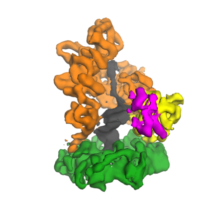 emd_4141.png emd_4141.png | 105.8 KB | ||
| Masks | emd_4141_msk_1.map | 10.5 MB | Mask map | |
| Filedesc metadata | emd-4141.cif.gz | 7.9 KB | ||
| Others | emd_4141_half_map_1.map.gzemd_4141_half_map_2.map.gz | 7.9 MB 7.9 MB | ||
| Archive directory |  http://ftp.pdbj.org/pub/emdb/structures/EMD-4141ftp://ftp.pdbj.org/pub/emdb/structures/EMD-4141 http://ftp.pdbj.org/pub/emdb/structures/EMD-4141ftp://ftp.pdbj.org/pub/emdb/structures/EMD-4141 | HTTPS FTP |
-Related structure data
| Related structure data | 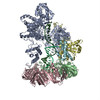 5m1sMC 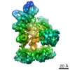 4142C M: atomic model generated by this map C: citing same article ( |
|---|---|
| Similar structure data |
-Links
| EMDB pages | EMDB (EBI/PDBe) / EMDataResource |
|---|---|
| Related items in Molecule of the Month |


-Map
| File | Download / File: emd_4141.map.gz / Format: CCP4 / Size: 10.5 MB / Type: IMAGE STORED AS FLOATING POINT NUMBER (4 BYTES) | ||||||||||||||||||||||||||||||||||||||||||||||||||||||||||||||||||||
|---|---|---|---|---|---|---|---|---|---|---|---|---|---|---|---|---|---|---|---|---|---|---|---|---|---|---|---|---|---|---|---|---|---|---|---|---|---|---|---|---|---|---|---|---|---|---|---|---|---|---|---|---|---|---|---|---|---|---|---|---|---|---|---|---|---|---|---|---|---|
| Annotation | Main map | ||||||||||||||||||||||||||||||||||||||||||||||||||||||||||||||||||||
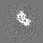
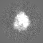
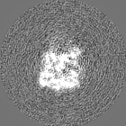
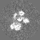
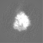
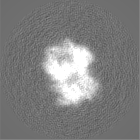
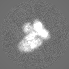
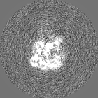
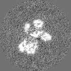
| Projections & slices | Image control
Images are generated by Spider. | ||||||||||||||||||||||||||||||||||||||||||||||||||||||||||||||||||||
| Voxel size | X=Y=Z: 1.76 Å | ||||||||||||||||||||||||||||||||||||||||||||||||||||||||||||||||||||
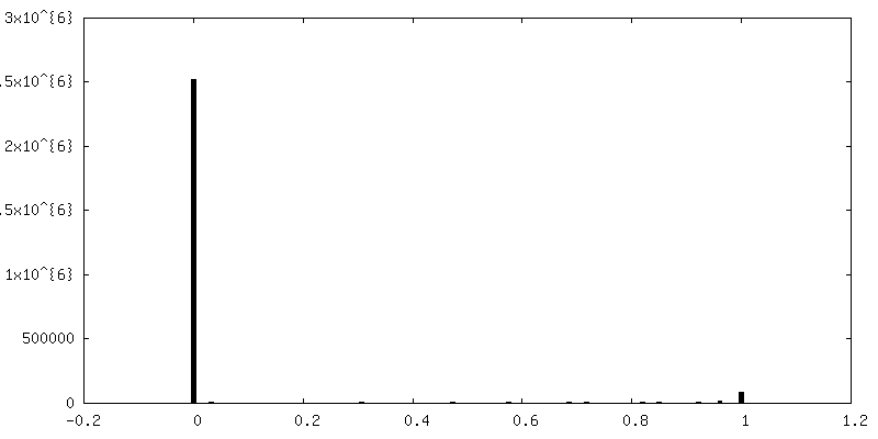
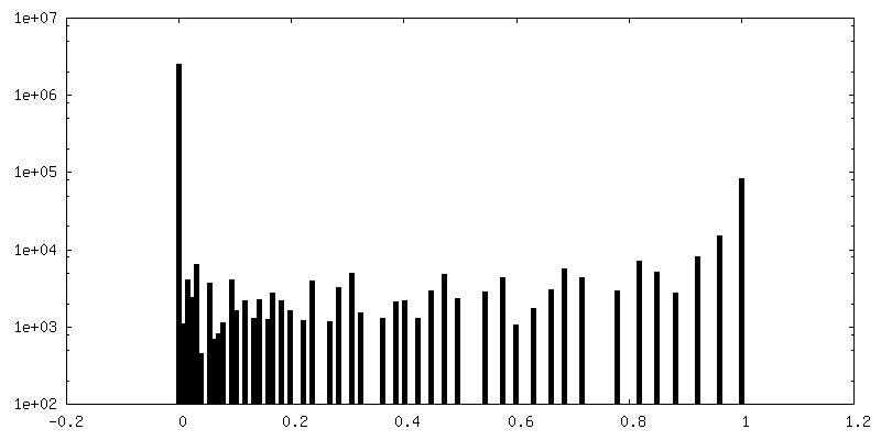
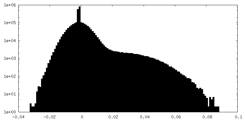
| Density |
| ||||||||||||||||||||||||||||||||||||||||||||||||||||||||||||||||||||
| Symmetry | Space group: 1 | ||||||||||||||||||||||||||||||||||||||||||||||||||||||||||||||||||||
| Details | EMDB XML:
CCP4 map header:
| ||||||||||||||||||||||||||||||||||||||||||||||||||||||||||||||||||||
 Z (Sec.)
 Y (Row.)
 X (Col.)


-Supplemental data
-Mask #1
| File | emd_4141_msk_1.map | ||||||||||||
|---|---|---|---|---|---|---|---|---|---|---|---|---|---|
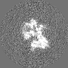
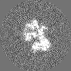
| Projections & Slices |
| ||||||||||||
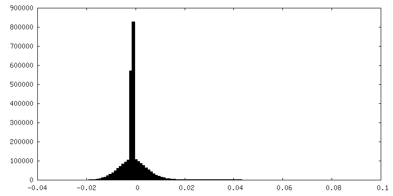
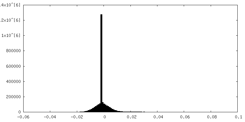
| Density Histograms |


-Half map: half map for main map
| File | emd_4141_half_map_1.map | ||||||||||||
|---|---|---|---|---|---|---|---|---|---|---|---|---|---|
| Annotation | half map for main map | ||||||||||||
| Projections & Slices |
| ||||||||||||
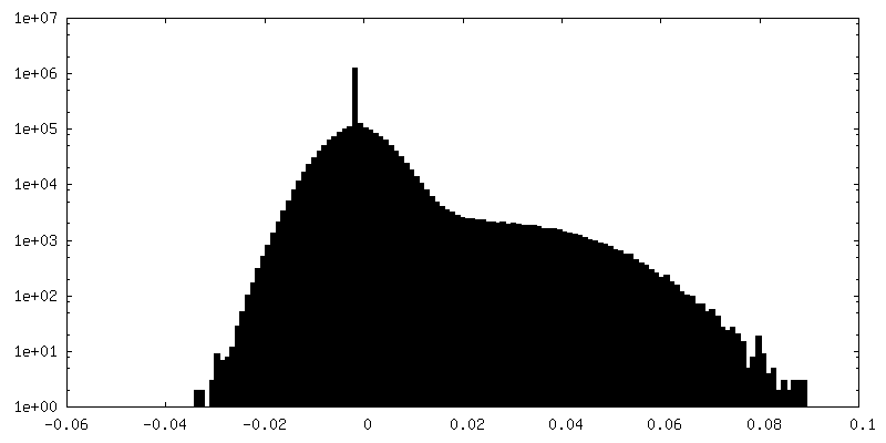
| Density Histograms |


-Half map: half map for main map
| File | emd_4141_half_map_2.map | ||||||||||||
|---|---|---|---|---|---|---|---|---|---|---|---|---|---|
| Annotation | half map for main map | ||||||||||||
| Projections & Slices |
| ||||||||||||
| Density Histograms |
- Sample components
-Entire : DNA polymerase III alpha, beta, epsilon, theta complex with misma...
| Entire | Name: DNA polymerase III alpha, beta, epsilon, theta complex with mismatched DNA duplex |
|---|---|
| Components |
|
-Supramolecule #1: DNA polymerase III alpha, beta, epsilon, theta complex with misma...
| Supramolecule | Name: DNA polymerase III alpha, beta, epsilon, theta complex with mismatched DNA duplex type: complex / ID: 1 / Parent: 0 / Macromolecule list: #1-#6 |
|---|---|
| Source (natural) | Organism: |
| Molecular weight | Theoretical: 250 KDa |
-Supramolecule #2: DNA polymerase III alpha, beta, epsilon, theta complex with misma...
| Supramolecule | Name: DNA polymerase III alpha, beta, epsilon, theta complex with mismatched DNA duplex type: complex / ID: 2 / Parent: 1 / Macromolecule list: #1-#3, #6 Details: Map obtained after signal subtraction of the beta subunit and alignment of the remaining parts. Final reconstruction obtained with non-subtracted images and angles from local alignment |
|---|---|
| Source (natural) | Organism: |
-Supramolecule #3: mismatched DNA duplex
| Supramolecule | Name: mismatched DNA duplex / type: complex / ID: 3 / Parent: 1 / Macromolecule list: #4-#5 |
|---|---|
| Source (natural) | Organism: synthetic construct (others) |
-Macromolecule #1: DNA polymerase III subunit alpha
| Macromolecule | Name: DNA polymerase III subunit alpha / type: protein_or_peptide / ID: 1 / Number of copies: 1 / Enantiomer: LEVO / EC number: DNA-directed DNA polymerase |
|---|---|
| Source (natural) | Organism: |
| Molecular weight | Theoretical: 103.554422 KDa |
| Recombinant expression | Organism: |
| Sequence | String: MSEPRFVHLR VHSDYSMIDG LAKTAPLVKK AAALGMPALA ITDFTNLCGL VKFYGAGHGA GIKPIVGADF NVQCDLLGDE LTHLTVLAA NNTGYQNLTL LISKAYQRGY GAAGPIIDRD WLIELNEGLI LLSGGRMGDV GRSLLRGNSA LVDECVAFYE E HFPDRYFL ...String: MSEPRFVHLR VHSDYSMIDG LAKTAPLVKK AAALGMPALA ITDFTNLCGL VKFYGAGHGA GIKPIVGADF NVQCDLLGDE LTHLTVLAA NNTGYQNLTL LISKAYQRGY GAAGPIIDRD WLIELNEGLI LLSGGRMGDV GRSLLRGNSA LVDECVAFYE E HFPDRYFL ELIRTGRPDE ESYLHAAVEL AEARGLPVVA TNDVRFIDSS DFDAHEIRVA IHDGFTLDDP KRPRNYSPQQ YM RSEEEMC ELFADIPEAL ANTVEIAKRC NVTVRLGEYF LPQFPTGDMS TEDYLVKRAK EGLEERLAFL FPDEEERLKR RPE YDERLE TELQVINQMG FPGYFLIVME FIQWSKDNGV PVGPGRGSGA GSLVAYALKI TDLDPLEFDL LFERFLNPER VSMP DFDVD FCMEKRDQVI EHVADMYGRD AVSQIITFGT MAAKAVIRDV GRVLGHPYGF VDRISKLIPP DPGMTLAKAF EAEPQ LPEI YEADEEVKAL IDMARKLEGV TRNAGKHAGG VVIAPTKITD FAPLYCDEEG KHPVTQFDKS DVEYAGLVKF DFLGLR TLT IINWALEMIN KRRAKNGEPP LDIAAIPLDD KKSFDMLQRS ETTAVFQLES RGMKDLIKRL QPDCFEDMIA LVALFRP GP LQSGMVDNFI DRKHGREEIS YPDVQWQHES LKPVLEPTYG IILYQEQVMQ IAQVLSGYTL GGADMLRRAM GKKKPEEM A KQRSVFAEGA EKNGINAELA MKIFDLVEKF AGYGFNKSHS AAYALVSYQT LWLKAHYPAE FMAAVMTADM DNTEKVVGL VDECWRMGLK ILPPDINSGL YHFHVNDDGE IVYGIGAIKG VGEGPIEAII EARNKGGYFR ELFDLCARTD TKKLNRRVLE KLIMSGAFD RLGPHRAALM NSLGDALKAA DQHAKAEAIG QLDLFGVL UniProtKB: DNA polymerase III subunit alpha |
-Macromolecule #2: DNA polymerase III subunit beta
| Macromolecule | Name: DNA polymerase III subunit beta / type: protein_or_peptide / ID: 2 / Number of copies: 2 / Enantiomer: LEVO / EC number: DNA-directed DNA polymerase |
|---|---|
| Source (natural) | Organism: |
| Molecular weight | Theoretical: 40.630508 KDa |
| Recombinant expression | Organism: |
| Sequence | String: MKFTVEREHL LKPLQQVSGP LGGRPTLPIL GNLLLQVADG TLSLTGTDLE MEMVARVALV QPHEPGATTV PARKFFDICR GLPEGAEIA VQLEGERMLV RSGRSRFSLS TLPAADFPNL DDWQSEVEFT LPQATMKRLI EATQFSMAHQ DVRYYLNGML F ETEGEELR ...String: MKFTVEREHL LKPLQQVSGP LGGRPTLPIL GNLLLQVADG TLSLTGTDLE MEMVARVALV QPHEPGATTV PARKFFDICR GLPEGAEIA VQLEGERMLV RSGRSRFSLS TLPAADFPNL DDWQSEVEFT LPQATMKRLI EATQFSMAHQ DVRYYLNGML F ETEGEELR TVATDGHRLA VCSMPIGQSL PSHSVIVPRK GVIELMRMLD GGDNPLRVQI GSNNIRAHVG DFIFTSKLVD GR FPDYRRV LPKNPDKHLE AGCDLLKQAF ARAAILSNEK FRGVRLYVSE NQLKITANNP EQEEAEEILD VTYSGAEMEI GFN VSYVLD VLNALKCENV RMMLTDSVSS VQIEDAASQS AAYVVMPMRL UniProtKB: Beta sliding clamp |
-Macromolecule #3: DNA polymerase III subunit epsilon
| Macromolecule | Name: DNA polymerase III subunit epsilon / type: protein_or_peptide / ID: 3 / Number of copies: 1 / Enantiomer: LEVO / EC number: DNA-directed DNA polymerase |
|---|---|
| Source (natural) | Organism: |
| Molecular weight | Theoretical: 27.118984 KDa |
| Recombinant expression | Organism: |
| Sequence | String: MSTAITRQIV LDTETTGMNQ IGAHYEGHKI IEIGAVEVVN RRLTGNNFHV YLKPDRLVDP EAFGVHGIAD EFLLDKPTFA EVADEFMDY IRGAELVIHN AAFDIGFMDY EFSLLKRDIP KTNTFCKVTD SLAVARKMFP GKRNSLDALC ARYEIDNSKR T LHGALLDA ...String: MSTAITRQIV LDTETTGMNQ IGAHYEGHKI IEIGAVEVVN RRLTGNNFHV YLKPDRLVDP EAFGVHGIAD EFLLDKPTFA EVADEFMDY IRGAELVIHN AAFDIGFMDY EFSLLKRDIP KTNTFCKVTD SLAVARKMFP GKRNSLDALC ARYEIDNSKR T LHGALLDA QILAEVYLAM TGGQLSLPLA MEGETQQQQG EATIQRIVRQ ASKLRVVFAT DEEIAAHEAR LDLVQKKGGS CL WRA UniProtKB: DNA polymerase III subunit epsilon |
-Macromolecule #6: DNA polymerase III subunit theta
| Macromolecule | Name: DNA polymerase III subunit theta / type: protein_or_peptide / ID: 6 / Number of copies: 1 / Enantiomer: LEVO / EC number: DNA-directed DNA polymerase |
|---|---|
| Source (natural) | Organism: |
| Molecular weight | Theoretical: 6.503385 KDa |
| Recombinant expression | Organism: |
| Sequence | String: QTEMDKVNVD LAAAGVAFKE RYNMPVIAEA VEREQPEHLR SWFRERLIAH RLASVN UniProtKB: DNA polymerase III subunit theta |
-Macromolecule #4: DNA Primer Strand
| Macromolecule | Name: DNA Primer Strand / type: dna / ID: 4 / Number of copies: 1 / Classification: DNA |
|---|---|
| Source (natural) | Organism: synthetic construct (others) |
| Molecular weight | Theoretical: 5.275448 KDa |
| Sequence | String: (DT)(DA)(DG)(DT)(DA)(DC)(DT)(DA)(DG)(DG) (DA)(DC)(DG)(DA)(DA)(DG)(DT) |
-Macromolecule #5: DNA Template Strand
| Macromolecule | Name: DNA Template Strand / type: dna / ID: 5 / Number of copies: 1 / Classification: DNA |
|---|---|
| Source (natural) | Organism: synthetic construct (others) |
| Molecular weight | Theoretical: 6.718339 KDa |
| Sequence | String: (DG)(DG)(DA)(DG)(DT)(DC)(DC)(DT)(DT)(DC) (DG)(DT)(DC)(DC)(DT)(DA)(DG)(DT)(DA)(DC) (DT)(DA) |
-Experimental details
-Structure determination
| Method | cryo EM |
|---|---|
 Processing Processing | single particle reconstruction |
| Aggregation state | particle |
-Sample preparation
| Concentration | 0.25 mg/mL | ||||||||||
|---|---|---|---|---|---|---|---|---|---|---|---|
| Buffer | pH: 7.5 Component:
| ||||||||||
| Grid | Model: Quantifoil R1.2/1.3 / Material: COPPER / Mesh: 300 / Pretreatment - Type: GLOW DISCHARGE / Pretreatment - Time: 60 sec. / Pretreatment - Atmosphere: AIR | ||||||||||
| Vitrification | Cryogen name: ETHANE / Chamber humidity: 100 % / Chamber temperature: 277 K / Instrument: FEI VITROBOT MARK IV Details: Prior to sample preparation 0.1 volumes of 0.05% Tween 20 were added to the sample 3 microliters were pipetted onto the grid and blotted for 4 seconds. | ||||||||||
| Details | Sample was run over a gel filtration column prior to vitrification |
- Electron microscopy
| Microscope | FEI TITAN KRIOS |
|---|---|
| Temperature | Min: 80.0 K / Max: 80.0 K |
| Specialist optics | Energy filter - Name: GIF Quantum / Energy filter - Lower energy threshold: 0 eV / Energy filter - Upper energy threshold: 20 eV |
| Image recording | Film or detector model: GATAN K2 SUMMIT (4k x 4k) / Detector mode: COUNTING / Digitization - Dimensions - Width: 3710 pixel / Digitization - Dimensions - Height: 3710 pixel / Digitization - Frames/image: 1-20 / Number grids imaged: 3 / Number real images: 1157 / Average exposure time: 25.0 sec. / Average electron dose: 2.0 e/Å2 |
| Electron beam | Acceleration voltage: 300 kV / Electron source:  FIELD EMISSION GUN FIELD EMISSION GUN |
| Electron optics | C2 aperture diameter: 50.0 µm / Calibrated magnification: 79545 / Illumination mode: FLOOD BEAM / Imaging mode: BRIGHT FIELD / Cs: 2.7 mm / Nominal defocus max: 3.5 µm / Nominal defocus min: 1.8 µm / Nominal magnification: 64000 |
| Sample stage | Specimen holder model: FEI TITAN KRIOS AUTOGRID HOLDER / Cooling holder cryogen: NITROGEN |
| Experimental equipment |  Model: Titan Krios / Image courtesy: FEI Company |
+Image processing
-Atomic model buiding 1
| Details | The cryo-EM structure of the PolIIIalpha-clamp-exonuclease complex in the polymerase mode (PDB code: 5FKW) was used as a starting model, and the NMR structure of theta bound to the exonuclease catalytic domain (PDB code: 2XY8) was used to place theta into the cryo-EM map. The model was manually adjusted in Coot and geometry of the protein optimized in Refmac5 using DNA-specific restraints generated in LibG |
|---|---|
| Refinement | Protocol: OTHER |
| Output model | PDB-5m1s: |
