 Movie
Movie Controller

[English] 日本語
 Yorodumi
- EMDB-4075: Structure of bacterial 30S-IF1-IF3-mRNA translation pre-initiatio... -
+ Open data

- Basic information
| Entry | Database: EMDB / ID: EMD-4075 | |||||||||
|---|---|---|---|---|---|---|---|---|---|---|
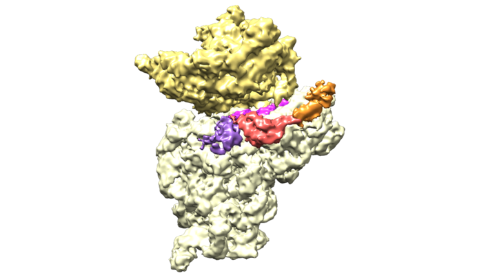
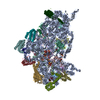
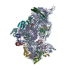
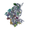
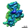
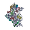
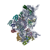
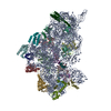
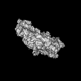
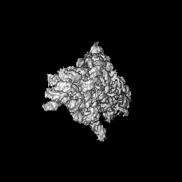
| Title | Structure of bacterial 30S-IF1-IF3-mRNA translation pre-initiation complex (state-1C) | |||||||||
 Map data Map data | For optimal visualization of IF3 N-terminal domain, gauss-filter de map by 1.1 and display it at 0.035 contour | |||||||||
 Sample Sample |
| |||||||||
 Keywords Keywords | ribosome / translation / initiation factors / 30S / IF1 / IF3 / PIC / Thermus thermophilus | |||||||||
| Function / homology |  Function and homology information Function and homology informationribosome disassembly / translation initiation factor activity / ribosomal small subunit assembly / ribosome binding / ribosomal small subunit biogenesis / small ribosomal subunit / small ribosomal subunit rRNA binding / cytosolic small ribosomal subunit / tRNA binding / rRNA binding ...ribosome disassembly / translation initiation factor activity / ribosomal small subunit assembly / ribosome binding / ribosomal small subunit biogenesis / small ribosomal subunit / small ribosomal subunit rRNA binding / cytosolic small ribosomal subunit / tRNA binding / rRNA binding / structural constituent of ribosome / ribosome / translation / ribonucleoprotein complex / mRNA binding / zinc ion binding / membrane / metal ion binding / cytosol / cytoplasm Similarity search - Function | |||||||||
| Biological species |   Thermus thermophilus HB8 (bacteria) / Thermus thermophilus (bacteria) / Thermus thermophilus (strain HB8 / ATCC 27634 / DSM 579) (bacteria) Thermus thermophilus HB8 (bacteria) / Thermus thermophilus (bacteria) / Thermus thermophilus (strain HB8 / ATCC 27634 / DSM 579) (bacteria) | |||||||||
| Method | single particle reconstruction / cryo EM / Resolution: 5.35 Å | |||||||||
 Authors Authors | Hussain T / Llacer JL | |||||||||
 Citation Citation | Journal: Cell / Year: 2016 Title: Large-Scale Movements of IF3 and tRNA during Bacterial Translation Initiation. Authors: Tanweer Hussain / Jose L Llácer / Brian T Wimberly / Jeffrey S Kieft / V Ramakrishnan /   Abstract: In bacterial translational initiation, three initiation factors (IFs 1-3) enable the selection of initiator tRNA and the start codon in the P site of the 30S ribosomal subunit. Here, we report 11 ...In bacterial translational initiation, three initiation factors (IFs 1-3) enable the selection of initiator tRNA and the start codon in the P site of the 30S ribosomal subunit. Here, we report 11 single-particle cryo-electron microscopy (cryoEM) reconstructions of the complex of bacterial 30S subunit with initiator tRNA, mRNA, and IFs 1-3, representing different steps along the initiation pathway. IF1 provides key anchoring points for IF2 and IF3, thereby enhancing their activities. IF2 positions a domain in an extended conformation appropriate for capturing the formylmethionyl moiety charged on tRNA. IF3 and tRNA undergo large conformational changes to facilitate the accommodation of the formylmethionyl-tRNA (fMet-tRNA(fMet)) into the P site for start codon recognition. | |||||||||
| History |
|
- Structure visualization
| Movie |
Movie viewer |
|---|---|
| Structure viewer | EM map: SurfViewMolmilJmol/JSmol |
| Supplemental images |
- Downloads & links
-EMDB archive
| Map data | emd_4075.map.gz | 59.6 MB | EMDB map data format | |
|---|---|---|---|---|
| Header (meta data) | emd-4075-v30.xmlemd-4075.xml | 43.6 KB 43.6 KB | Display Display | EMDB header |
| Images |  emd_4075.png emd_4075.png | 167.4 KB | ||
| Filedesc metadata | emd-4075.cif.gz | 9.3 KB | ||
| Others | emd_4075_half_map_1.map.gzemd_4075_half_map_2.map.gz | 57.4 MB 58 MB | ||
| Archive directory |  http://ftp.pdbj.org/pub/emdb/structures/EMD-4075ftp://ftp.pdbj.org/pub/emdb/structures/EMD-4075 http://ftp.pdbj.org/pub/emdb/structures/EMD-4075ftp://ftp.pdbj.org/pub/emdb/structures/EMD-4075 | HTTPS FTP |
-Related structure data
| Related structure data |  5lmpMC  4073C  4074C 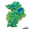 4076C 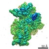 4077C 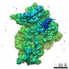 4078C 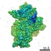 4079C 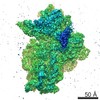 4080C 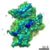 4081C 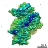 4082C  4083C 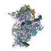 5lmnC 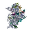 5lmoC 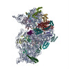 5lmqC  5lmrC 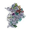 5lmsC 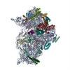 5lmtC 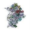 5lmuC 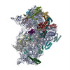 5lmvC M: atomic model generated by this map C: citing same article ( |
|---|---|
| Similar structure data |


-Links
| EMDB pages | EMDB (EBI/PDBe) / EMDataResource |
|---|---|
| Related items in Molecule of the Month |


-Map
| File | Download / File: emd_4075.map.gz / Format: CCP4 / Size: 67 MB / Type: IMAGE STORED AS FLOATING POINT NUMBER (4 BYTES) | ||||||||||||||||||||||||||||||||||||||||||||||||||||||||||||
|---|---|---|---|---|---|---|---|---|---|---|---|---|---|---|---|---|---|---|---|---|---|---|---|---|---|---|---|---|---|---|---|---|---|---|---|---|---|---|---|---|---|---|---|---|---|---|---|---|---|---|---|---|---|---|---|---|---|---|---|---|---|
| Annotation | For optimal visualization of IF3 N-terminal domain, gauss-filter de map by 1.1 and display it at 0.035 contour | ||||||||||||||||||||||||||||||||||||||||||||||||||||||||||||
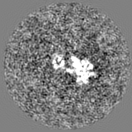
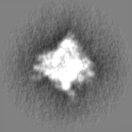
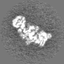
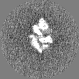
| Projections & slices | Image control
Images are generated by Spider. | ||||||||||||||||||||||||||||||||||||||||||||||||||||||||||||
| Voxel size | X=Y=Z: 1.34 Å | ||||||||||||||||||||||||||||||||||||||||||||||||||||||||||||
| Density |
| ||||||||||||||||||||||||||||||||||||||||||||||||||||||||||||
| Symmetry | Space group: 1 | ||||||||||||||||||||||||||||||||||||||||||||||||||||||||||||
| Details | EMDB XML:
CCP4 map header:
| ||||||||||||||||||||||||||||||||||||||||||||||||||||||||||||
 Z (Sec.)
 Y (Row.)
 X (Col.)


-Supplemental data
-Half map: #1
| File | emd_4075_half_map_1.map | ||||||||||||
|---|---|---|---|---|---|---|---|---|---|---|---|---|---|
| Projections & Slices |
| ||||||||||||
| Density Histograms |


-Half map: #2
| File | emd_4075_half_map_2.map | ||||||||||||
|---|---|---|---|---|---|---|---|---|---|---|---|---|---|
| Projections & Slices |
| ||||||||||||
| Density Histograms |


- Sample components
+Entire : 30S-IF1-IF3-mRNA pre-initiation complex (state-1C)
+Supramolecule #1: 30S-IF1-IF3-mRNA pre-initiation complex (state-1C)
+Macromolecule #1: 16S rRNA
+Macromolecule #24: mRNA
+Macromolecule #2: 30S ribosomal protein S2
+Macromolecule #3: 30S ribosomal protein S3
+Macromolecule #4: 30S ribosomal protein S4
+Macromolecule #5: 30S ribosomal protein S5
+Macromolecule #6: 30S ribosomal protein S6
+Macromolecule #7: 30S ribosomal protein S7
+Macromolecule #8: 30S ribosomal protein S8
+Macromolecule #9: 30S ribosomal protein S9
+Macromolecule #10: 30S ribosomal protein S10
+Macromolecule #11: 30S ribosomal protein S11
+Macromolecule #12: 30S ribosomal protein S12
+Macromolecule #13: 30S ribosomal protein S13
+Macromolecule #14: 30S ribosomal protein S14 type Z
+Macromolecule #15: 30S ribosomal protein S15
+Macromolecule #16: 30S ribosomal protein S16
+Macromolecule #17: 30S ribosomal protein S17
+Macromolecule #18: 30S ribosomal protein S18
+Macromolecule #19: 30S ribosomal protein S19
+Macromolecule #20: 30S ribosomal protein S20
+Macromolecule #21: 30S ribosomal protein Thx
+Macromolecule #22: Translation initiation factor IF-1
+Macromolecule #23: Translation initiation factor IF-3
+Macromolecule #25: MAGNESIUM ION
+Macromolecule #26: ZINC ION
-Experimental details
-Structure determination
| Method | cryo EM |
|---|---|
 Processing Processing | single particle reconstruction |
| Aggregation state | particle |
-Sample preparation
| Concentration | 0.08 mg/mL | ||||||||||||
|---|---|---|---|---|---|---|---|---|---|---|---|---|---|
| Buffer | pH: 7.5 Component:
| ||||||||||||
| Grid | Model: Quantifoil / Material: COPPER / Mesh: 400 / Support film - Material: CARBON / Support film - topology: HOLEY / Pretreatment - Type: GLOW DISCHARGE / Pretreatment - Time: 20 sec. | ||||||||||||
| Vitrification | Cryogen name: ETHANE / Chamber humidity: 100 % / Chamber temperature: 277 K / Instrument: FEI VITROBOT MARK I |
- Electron microscopy
| Microscope | FEI POLARA 300 |
|---|---|
| Temperature | Min: 90.0 K / Max: 100.0 K |
| Image recording | Film or detector model: OTHER / Number grids imaged: 5 / Number real images: 4400 / Average exposure time: 1.1 sec. / Average electron dose: 30.0 e/Å2 Details: Recorded in a FEI Falcon III direct electron detector. Images were collected in movie-mode at 32 frames per second |
| Electron beam | Acceleration voltage: 300 kV / Electron source:  FIELD EMISSION GUN FIELD EMISSION GUN |
| Electron optics | C2 aperture diameter: 50.0 µm / Calibrated magnification: 104478 / Illumination mode: FLOOD BEAM / Imaging mode: BRIGHT FIELD / Cs: 2.0 mm / Nominal defocus max: 3.5 µm / Nominal defocus min: 1.5 µm / Nominal magnification: 78000 |
| Sample stage | Cooling holder cryogen: NITROGEN |
| Experimental equipment |  Model: Tecnai Polara / Image courtesy: FEI Company |
+Image processing

-Atomic model buiding 1
| Refinement | Space: RECIPROCAL / Protocol: OTHER / Target criteria: FSC |
|---|---|
| Output model | PDB-5lmp: |
