 ムービー
ムービー コントローラー


+ データを開く

- 基本情報
基本情報
| 登録情報 | データベース: PDB / ID: 3jbs | ||||||
|---|---|---|---|---|---|---|---|
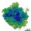
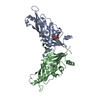
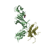
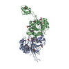
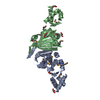
| タイトル | eL6 protein from yeast 60S ribosomal subunit | ||||||
 要素 要素 | eL6 | ||||||
 キーワード キーワード | TRANSLATION / ribosome | ||||||
| 機能・相同性 |  機能・相同性情報 機能・相同性情報SRP-dependent cotranslational protein targeting to membrane / GTP hydrolysis and joining of the 60S ribosomal subunit / Formation of a pool of free 40S subunits / Nonsense Mediated Decay (NMD) independent of the Exon Junction Complex (EJC) / Nonsense Mediated Decay (NMD) enhanced by the Exon Junction Complex (EJC) / L13a-mediated translational silencing of Ceruloplasmin expression / ribosomal large subunit assembly / cytosolic large ribosomal subunit / cytoplasmic translation / structural constituent of ribosome ...SRP-dependent cotranslational protein targeting to membrane / GTP hydrolysis and joining of the 60S ribosomal subunit / Formation of a pool of free 40S subunits / Nonsense Mediated Decay (NMD) independent of the Exon Junction Complex (EJC) / Nonsense Mediated Decay (NMD) enhanced by the Exon Junction Complex (EJC) / L13a-mediated translational silencing of Ceruloplasmin expression / ribosomal large subunit assembly / cytosolic large ribosomal subunit / cytoplasmic translation / structural constituent of ribosome / RNA binding / cytosol 類似検索 - 分子機能 | ||||||
| 生物種 |  | ||||||
| 手法 | 電子顕微鏡法 / 単粒子再構成法 / クライオ電子顕微鏡法 / 解像度: 2.9 Å | ||||||
 データ登録者 データ登録者 | Passos, D.O. / Lyumkis, D. | ||||||
 引用 引用 | ジャーナル: J Struct Biol / 年: 2015 タイトル: Single-particle cryoEM analysis at near-atomic resolution from several thousand asymmetric subunits. 著者: Dario Oliveira Passos / Dmitry Lyumkis /  要旨: A single-particle cryoEM reconstruction of the large ribosomal subunit from Saccharomyces cerevisiae was obtained from a dataset of ∼75,000 particles. The gold-standard and frequency-limited ...A single-particle cryoEM reconstruction of the large ribosomal subunit from Saccharomyces cerevisiae was obtained from a dataset of ∼75,000 particles. The gold-standard and frequency-limited approaches to single-particle refinement were each independently used to determine orientation parameters for the final reconstruction. Both approaches showed similar resolution curves and nominal resolution values for the 60S dataset, estimated at 2.9 Å. The amount of over-fitting present during frequency-limited refinement was quantitatively analyzed using the high-resolution phase-randomization test, and the results showed no apparent over-fitting. The number of asymmetric subunits required to reach specific resolutions was subsequently analyzed by refining subsets of the data in an ab initio manner. With our data collection and processing strategies, sub-nanometer resolution was obtained with ∼200 asymmetric subunits (or, equivalently for the ribosomal subunit, particles). Resolutions of 5.6 Å, 4.5 Å, and 3.8 Å were reached with ∼1000, ∼1600, and ∼5000 asymmetric subunits, respectively. At these resolutions, one would expect to detect alpha-helical pitch, separation of beta-strands, and separation of Cα atoms, respectively. Using this map, together with strategies for ab initio model building and model refinement, we built a region of the ribosomal protein eL6, which was missing in previous models of the yeast ribosome. The relevance for more routine high-resolution structure determination is discussed. | ||||||
| 履歴 |
|
- 構造の表示
構造の表示
| ムービー |
ムービービューア |
|---|---|
| 構造ビューア | 分子: MolmilJmol/JSmol |
- ダウンロードとリンク
-ダウンロード
| PDBx/mmCIF形式 | 3jbs.cif.gz | 45.1 KB | 表示 | PDBx/mmCIF形式 |
|---|---|---|---|---|
| PDB形式 | pdb3jbs.ent.gz | 31.8 KB | 表示 | PDB形式 |
| PDBx/mmJSON形式 | 3jbs.json.gz | ツリー表示 | PDBx/mmJSON形式 | |
| その他 |  その他のダウンロード その他のダウンロード |
-検証レポート
| 文書・要旨 | 3jbs_validation.pdf.gz | 819.9 KB | 表示 | wwPDB検証レポート |
|---|---|---|---|---|
| 文書・詳細版 | 3jbs_full_validation.pdf.gz | 819.5 KB | 表示 | |
| XML形式データ | 3jbs_validation.xml.gz | 12.6 KB | 表示 | |
| CIF形式データ | 3jbs_validation.cif.gz | 16.8 KB | 表示 | |
| アーカイブディレクトリ | https://data.pdbj.org/pub/pdb/validation_reports/jb/3jbsftp://data.pdbj.org/pub/pdb/validation_reports/jb/3jbs | HTTPS FTP |
-関連構造データ


-リンク
 PDBj
PDBj


- 集合体
集合体
| 登録構造単位 | 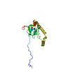
|
|---|---|
| 1 |
|
-要素
| #1: タンパク質 | 分子量: 20000.564 Da / 分子数: 1 / 由来タイプ: 天然 / 詳細: cytosolic extract 由来: (天然) 株: BY4741 / 参照: UniProt: Q02326 |
|---|
-実験情報
-実験
| 実験 | 手法: 電子顕微鏡法 |
|---|---|
| EM実験 | 試料の集合状態: PARTICLE / 3次元再構成法: 単粒子再構成法 |
- 試料調製
試料調製
| 構成要素 | 名称: Saccharomyces cerevisiae large 60S ribosomal subunit タイプ: RIBOSOME |
|---|---|
| 分子量 | 値: 2.5 MDa / 実験値: NO |
| 緩衝液 | 名称: elution buffer (50 mM HEPES-KOH, 100 mM KOAc, 5 mM MgOAc, 1 mM EDTA, 2 mM DTT) pH: 6.8 詳細: elution buffer (50 mM HEPES-KOH, 100 mM KOAc, 5 mM MgOAc, 1 mM EDTA, 2 mM DTT) |
| 試料 | 包埋: NO / シャドウイング: NO / 染色: NO / 凍結: YES |
| 試料支持 | 詳細: 400 mesh, 1.2x1.3 micron C-flat grids, plasma-treated for 5 seconds |
| 急速凍結 | 装置: HOMEMADE PLUNGER / 凍結剤: ETHANE / Temp: 77 K / 湿度: 90 % 詳細: A 3 uL sample was applied onto a freshly plasma-treated (6 seconds, Gatan Solarus plasma cleaner) holey carbon C-flat grid (Protochips, Inc.), allowing the sample to adsorb for 30 seconds. ...詳細: A 3 uL sample was applied onto a freshly plasma-treated (6 seconds, Gatan Solarus plasma cleaner) holey carbon C-flat grid (Protochips, Inc.), allowing the sample to adsorb for 30 seconds. The sample was then plunge-frozen in liquid ethane using a manual cryo-plunger in ambient atmosphere at 4 degrees C. 手法: A 3 uL sample was applied onto a freshly plasma-treated (6 seconds, Gatan Solarus plasma cleaner) holey carbon C-flat grid (Protochips, Inc.), allowing the sample to adsorb for 30 seconds. ...手法: A 3 uL sample was applied onto a freshly plasma-treated (6 seconds, Gatan Solarus plasma cleaner) holey carbon C-flat grid (Protochips, Inc.), allowing the sample to adsorb for 30 seconds. The sample was then plunge-frozen in liquid ethane using a manual cryo-plunger in ambient atmosphere at 4 degrees C. |
- 電子顕微鏡撮影
| 実験機器 |  モデル: Titan Krios / 画像提供: FEI Company |
|---|---|
| 顕微鏡 | モデル: FEI TITAN KRIOS / 日付: 2014年9月2日 / 詳細: super-resolution imaging |
| 電子銃 | 電子線源:  FIELD EMISSION GUN / 加速電圧: 300 kV / 照射モード: FLOOD BEAM FIELD EMISSION GUN / 加速電圧: 300 kV / 照射モード: FLOOD BEAM |
| 電子レンズ | モード: BRIGHT FIELD / 倍率(公称値): 22500 X / 倍率(補正後): 38167 X / 最大 デフォーカス(公称値): 2500 nm / 最小 デフォーカス(公称値): 500 nm / Cs: 2.7 mm 非点収差: Objective lens astigmatism was corrected within the Leginon software. |
| 試料ホルダ | 試料ホルダーモデル: FEI TITAN KRIOS AUTOGRID HOLDER |
| 撮影 | 電子線照射量: 25 e/Å2 / フィルム・検出器のモデル: GATAN K2 (4k x 4k) |
| 画像スキャン | デジタル画像の数: 1833 |
- 解析
解析
| EMソフトウェア |
| ||||||||||||
|---|---|---|---|---|---|---|---|---|---|---|---|---|---|
| CTF補正 | 詳細: each particle | ||||||||||||
| 対称性 | 点対称性: C1 (非対称) | ||||||||||||
| 3次元再構成 | 手法: projection matching / 解像度: 2.9 Å / 解像度の算出法: FSC 0.143 CUT-OFF / 粒子像の数: 75653 / ピクセルサイズ(公称値): 1.31 Å / ピクセルサイズ(実測値): 1.31 Å 詳細: B-factor of -50 applied to final map (Single particle--Applied symmetry: C1). 対称性のタイプ: POINT | ||||||||||||
| 原子モデル構築 | B value: 50 / プロトコル: FLEXIBLE FIT / 空間: REAL / Target criteria: FSC / 詳細: REFINEMENT PROTOCOL--flexible | ||||||||||||
| 原子モデル構築 | PDB-ID: 4UJQ 4ujq PDB chain-ID: H / Accession code: 4UJQ / Source name: PDB / タイプ: experimental model | ||||||||||||
| 精密化ステップ | サイクル: LAST
|
