[English] 日本語
 Yorodumi
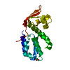
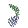
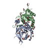
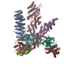
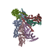
- PDB-3j8c: Model of the human eIF3 PCI-MPN octamer docked into the 43S EM map -
+ Open data
- Basic information
| Entry | Database: PDB / ID: 3j8c | |||||||||
|---|---|---|---|---|---|---|---|---|---|---|
| Title | Model of the human eIF3 PCI-MPN octamer docked into the 43S EM map | |||||||||
 Components Components | (Eukaryotic translation initiation factor 3 subunit ...) x 8 | |||||||||
 Keywords Keywords | TRANSLATION | |||||||||
| Function / homology |  Function and homology information Function and homology informationpositive regulation of mRNA binding / viral translational termination-reinitiation / eukaryotic translation initiation factor 3 complex, eIF3e / eukaryotic translation initiation factor 3 complex, eIF3m / IRES-dependent viral translational initiation / translation reinitiation / formation of cytoplasmic translation initiation complex / cytoplasmic translational initiation / multi-eIF complex / eukaryotic translation initiation factor 3 complex ...positive regulation of mRNA binding / viral translational termination-reinitiation / eukaryotic translation initiation factor 3 complex, eIF3e / eukaryotic translation initiation factor 3 complex, eIF3m / IRES-dependent viral translational initiation / translation reinitiation / formation of cytoplasmic translation initiation complex / cytoplasmic translational initiation / multi-eIF complex / eukaryotic translation initiation factor 3 complex / eukaryotic 43S preinitiation complex / eukaryotic 48S preinitiation complex / nuclear-transcribed mRNA catabolic process, nonsense-mediated decay / metal-dependent deubiquitinase activity / regulation of translational initiation / Formation of the ternary complex, and subsequently, the 43S complex / Ribosomal scanning and start codon recognition / Translation initiation complex formation / Formation of a pool of free 40S subunits / GTP hydrolysis and joining of the 60S ribosomal subunit / L13a-mediated translational silencing of Ceruloplasmin expression / translation initiation factor binding / negative regulation of translational initiation / translation initiation factor activity / negative regulation of proteasomal ubiquitin-dependent protein catabolic process / positive regulation of translation / translational initiation / PML body / receptor tyrosine kinase binding / negative regulation of ERK1 and ERK2 cascade / fibrillar center / metallopeptidase activity / ribosome binding / microtubule / ubiquitinyl hydrolase 1 / cysteine-type deubiquitinase activity / postsynaptic density / cadherin binding / mRNA binding / synapse / chromatin / nucleolus / structural molecule activity / proteolysis / RNA binding / extracellular exosome / nucleoplasm / identical protein binding / membrane / nucleus / cytoplasm / cytosol Similarity search - Function | |||||||||
| Biological species |  Homo sapiens (human) Homo sapiens (human) | |||||||||
| Method | ELECTRON MICROSCOPY / single particle reconstruction / cryo EM / Resolution: 11.6 Å | |||||||||
 Authors Authors | Erzberger, J.P. / Ban, N. | |||||||||
 Citation Citation |  Journal: Cell / Year: 2013 Journal: Cell / Year: 2013Title: Structure of the mammalian ribosomal 43S preinitiation complex bound to the scanning factor DHX29. Authors: Yaser Hashem / Amedee des Georges / Vidya Dhote / Robert Langlois / Hstau Y Liao / Robert A Grassucci / Christopher U T Hellen / Tatyana V Pestova / Joachim Frank /  Abstract: Eukaryotic translation initiation begins with assembly of a 43S preinitiation complex. First, methionylated initiator methionine transfer RNA (Met-tRNAi(Met)), eukaryotic initiation factor (eIF) 2, ...Eukaryotic translation initiation begins with assembly of a 43S preinitiation complex. First, methionylated initiator methionine transfer RNA (Met-tRNAi(Met)), eukaryotic initiation factor (eIF) 2, and guanosine triphosphate form a ternary complex (TC). The TC, eIF3, eIF1, and eIF1A cooperatively bind to the 40S subunit, yielding the 43S preinitiation complex, which is ready to attach to messenger RNA (mRNA) and start scanning to the initiation codon. Scanning on structured mRNAs additionally requires DHX29, a DExH-box protein that also binds directly to the 40S subunit. Here, we present a cryo-electron microscopy structure of the mammalian DHX29-bound 43S complex at 11.6 Å resolution. It reveals that eIF2 interacts with the 40S subunit via its α subunit and supports Met-tRNAi(Met) in an unexpected P/I orientation (eP/I). The structural core of eIF3 resides on the back of the 40S subunit, establishing two principal points of contact, whereas DHX29 binds around helix 16. The structure provides insights into eukaryote-specific aspects of translation, including the mechanism of action of DHX29. | |||||||||
| History |
| |||||||||
| Remark 0 | THIS ENTRY 3J8C CONTAINS A STRUCTURAL MODEL FIT TO AN ELECTRON MICROSCOPY MAP (EMD-5658) DETERMINED ...THIS ENTRY 3J8C CONTAINS A STRUCTURAL MODEL FIT TO AN ELECTRON MICROSCOPY MAP (EMD-5658) DETERMINED ORIGINALLY BY AUTHORS: Y.HASHEM, A.DES-GEORGES, V.DHOTE, R.LANGLOIS, H.Y.LIAO, R.A.GRASSUCCI, C.U.T.HELLEN, T.V.PESTOVA, J.FRANK | |||||||||
| Remark 650 | HELIX DETERMINATION METHOD: AUTHOR DETERMINED | |||||||||
| Remark 700 | SHEET DETERMINATION METHOD: AUTHOR DETERMINED |
- Structure visualization
| Movie |
 Movie viewer Movie viewer |
|---|---|
| Structure viewer | Molecule:  Molmil Molmil Jmol/JSmol Jmol/JSmol |
- Downloads & links
- Download
| PDBx/mmCIF format |  3j8c.cif.gz 3j8c.cif.gz | 368.2 KB | Display |  PDBx/mmCIF format PDBx/mmCIF format |
|---|---|---|---|---|
| PDB format |  pdb3j8c.ent.gz pdb3j8c.ent.gz | 223.3 KB | Display |  PDB format PDB format |
| PDBx/mmJSON format |  3j8c.json.gz 3j8c.json.gz | Tree view |  PDBx/mmJSON format PDBx/mmJSON format | |
| Others |  Other downloads Other downloads |
-Validation report
| Arichive directory |  https://data.pdbj.org/pub/pdb/validation_reports/j8/3j8c https://data.pdbj.org/pub/pdb/validation_reports/j8/3j8c ftp://data.pdbj.org/pub/pdb/validation_reports/j8/3j8c ftp://data.pdbj.org/pub/pdb/validation_reports/j8/3j8c | HTTPS FTP |
|---|
-Related structure data
| Related structure data |  5658M  2670C  2671C  3j8bC  4u1cC  4u1dC  4u1eC  4u1fC M: map data used to model this data C: citing same article ( |
|---|---|
| Similar structure data |
- Links
Links
- Assembly
| Deposited unit | 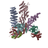
|
|---|---|
| 1 |
|
- Components
-Eukaryotic translation initiation factor 3 subunit ... , 8 types, 8 molecules ACEFHKLM
| #1: Protein | Mass: 61036.340 Da / Num. of mol.: 1 / Fragment: SEE REMARK 999 / Source method: isolated from a natural source / Source: (natural)  Homo sapiens (human) / References: UniProt: Q14152*PLUS Homo sapiens (human) / References: UniProt: Q14152*PLUS |
|---|---|
| #2: Protein | Mass: 62812.340 Da / Num. of mol.: 1 / Fragment: SEE REMARK 999 / Source method: isolated from a natural source / Source: (natural)  Homo sapiens (human) / References: UniProt: Q99613*PLUS Homo sapiens (human) / References: UniProt: Q99613*PLUS |
| #3: Protein | Mass: 48854.117 Da / Num. of mol.: 1 / Fragment: SEE REMARK 999 / Source method: isolated from a natural source / Source: (natural)  Homo sapiens (human) / References: UniProt: P60228*PLUS Homo sapiens (human) / References: UniProt: P60228*PLUS |
| #4: Protein | Mass: 32756.850 Da / Num. of mol.: 1 / Fragment: SEE REMARK 999 / Source method: isolated from a natural source / Source: (natural)  Homo sapiens (human) / References: UniProt: O00303*PLUS, ubiquitinyl hydrolase 1 Homo sapiens (human) / References: UniProt: O00303*PLUS, ubiquitinyl hydrolase 1 |
| #5: Protein | Mass: 35125.961 Da / Num. of mol.: 1 / Fragment: SEE REMARK 999 / Source method: isolated from a natural source / Source: (natural)  Homo sapiens (human) / References: UniProt: O15372*PLUS Homo sapiens (human) / References: UniProt: O15372*PLUS |
| #6: Protein | Mass: 23533.061 Da / Num. of mol.: 1 / Fragment: SEE REMARK 999 / Source method: isolated from a natural source / Source: (natural)  Homo sapiens (human) / References: UniProt: Q9UBQ5*PLUS Homo sapiens (human) / References: UniProt: Q9UBQ5*PLUS |
| #7: Protein | Mass: 63008.434 Da / Num. of mol.: 1 / Fragment: SEE REMARK 999 / Source method: isolated from a natural source / Source: (natural)  Homo sapiens (human) / References: UniProt: Q9Y262*PLUS Homo sapiens (human) / References: UniProt: Q9Y262*PLUS |
| #8: Protein | Mass: 40404.781 Da / Num. of mol.: 1 / Fragment: SEE REMARK 999 / Source method: isolated from a natural source / Source: (natural)  Homo sapiens (human) / References: UniProt: Q7L2H7*PLUS Homo sapiens (human) / References: UniProt: Q7L2H7*PLUS |
-Details
| Sequence details | ENTRY CONTAINS EIF3 PROTEINS FROM HOMO SAPIENS FITTED INTO ELECTRON MICROSCOPY DATA DERIVED FROM ...ENTRY CONTAINS EIF3 PROTEINS FROM HOMO SAPIENS FITTED INTO ELECTRON MICROSCOPY |
|---|
-Experimental details
-Experiment
| Experiment | Method: ELECTRON MICROSCOPY |
|---|---|
| EM experiment | Aggregation state: PARTICLE / 3D reconstruction method: single particle reconstruction |
- Sample preparation
| Component | Name: mammalian 43S preinitiation complex bound to DHX29 / Type: RIBOSOME |
|---|---|
| Buffer solution | pH: 7.5 |
| Specimen | Embedding applied: NO / Shadowing applied: NO / Staining applied: NO / Vitrification applied: YES |
| Vitrification | Instrument: FEI VITROBOT MARK II / Cryogen name: ETHANE |
- Electron microscopy imaging
| Microscopy | Model: FEI TECNAI 20 / Date: Nov 1, 2012 |
|---|---|
| Electron gun | Electron source:  FIELD EMISSION GUN / Accelerating voltage: 110 kV / Illumination mode: FLOOD BEAM FIELD EMISSION GUN / Accelerating voltage: 110 kV / Illumination mode: FLOOD BEAM |
| Electron lens | Mode: BRIGHT FIELD / Nominal defocus max: -4000 nm / Nominal defocus min: -1000 nm |
| Image recording | Electron dose: 12 e/Å2 / Film or detector model: GATAN ULTRASCAN 4000 (4k x 4k) |
| Radiation | Protocol: SINGLE WAVELENGTH / Monochromatic (M) / Laue (L): M / Scattering type: x-ray |
| Radiation wavelength | Relative weight: 1 |
- Processing
| EM software |
| ||||||||||||||||||||||||||||||||||||||||||||||||||
|---|---|---|---|---|---|---|---|---|---|---|---|---|---|---|---|---|---|---|---|---|---|---|---|---|---|---|---|---|---|---|---|---|---|---|---|---|---|---|---|---|---|---|---|---|---|---|---|---|---|---|---|
| Symmetry | Point symmetry: C1 (asymmetric) | ||||||||||||||||||||||||||||||||||||||||||||||||||
| 3D reconstruction | Resolution: 11.6 Å / Resolution method: FSC 0.143 CUT-OFF / Num. of particles: 29000 / Refinement type: HALF-MAPS REFINED INDEPENDENTLY / Symmetry type: POINT | ||||||||||||||||||||||||||||||||||||||||||||||||||
| Atomic model building | 3D fitting-ID: 1 / Source name: PDB / Type: experimental model
| ||||||||||||||||||||||||||||||||||||||||||||||||||
| Refinement step | Cycle: LAST
|
 Movie
Movie Controller






 PDBj
PDBj


