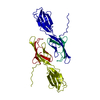
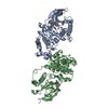
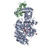
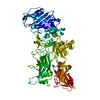
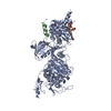
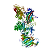
ジャーナル: Proc Natl Acad Sci U S A / 年: 2012 タイトル: Structural basis for TetM-mediated tetracycline resistance. 著者: Alexandra Dönhöfer / Sibylle Franckenberg / Stephan Wickles / Otto Berninghausen / Roland Beckmann / Daniel N Wilson / 要旨: Ribosome protection proteins (RPPs) confer tetracycline resistance by binding to the ribosome and chasing the drug from its binding site. The current model for the mechanism of action of RPPs ...Ribosome protection proteins (RPPs) confer tetracycline resistance by binding to the ribosome and chasing the drug from its binding site. The current model for the mechanism of action of RPPs proposes that drug release is indirect and achieved via conformational changes within the drug-binding site induced upon binding of the RPP to the ribosome. Here we report a cryo-EM structure of the RPP TetM in complex with the 70S ribosome at 7.2-Å resolution. The structure reveals the contacts of TetM with the ribosome, including interaction between the conserved and functionally critical C-terminal extension of TetM and the decoding center of the small subunit. Moreover, we observe direct interaction between domain IV of TetM and the tetracycline binding site and identify residues critical for conferring tetracycline resistance. A model is presented whereby TetM directly dislodges tetracycline to confer resistance.
モード: BRIGHT FIELD / 倍率(公称値): 75000 X / 最大 デフォーカス(公称値): -3500 nm / 最小 デフォーカス(公称値): -1000 nm
撮影
電子線照射量: 20 e/Å2 フィルム・検出器のモデル: TVIPS TEMCAM-F416 (4k x 4k)
放射
プロトコル: SINGLE WAVELENGTH / 単色(M)・ラウエ(L): M / 散乱光タイプ: x-ray
放射波長
相対比: 1
-
解析
EMソフトウェア
ID
名称
カテゴリ
1
Coot
モデルフィッティング
2
DireX
モデルフィッティング
3
UCSF Chimera
モデルフィッティング
4
SPIDER
3次元再構成
CTF補正
詳細: The volumes were CTF-corrected in defocus groups
対称性
点対称性: C1 (非対称)
3次元再構成
手法: back-projection interpolated in Fourier space / 解像度: 7.2 Å / 粒子像の数: 52701 / ピクセルサイズ(公称値): 1.04 Å / ピクセルサイズ(実測値): 1.04 Å 詳細: a modified version of SPIDER program was used for the reconstruction 対称性のタイプ: POINT
原子モデル構築
プロトコル: FLEXIBLE FIT / 空間: REAL / Target criteria: Cross-correlation coefficient 詳細: METHOD--Local refinement, Flexible fitting REFINEMENT PROTOCOL--rigid body
 ムービー
ムービー コントローラー


 データを開く
 基本情報
基本情報 要素
要素 キーワード
キーワード 機能・相同性情報
 Enterococcus faecalis (乳酸球菌)
 データ登録者
 引用
引用
 構造の表示
構造の表示 ダウンロードとリンク
 その他のダウンロード


 PDBj
PDBj


 集合体
集合体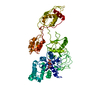

 分子量: 521.208 Da / 分子数: 1 / 由来タイプ: 合成 / 式: C11H18N5O13P3
 試料調製
試料調製 電子顕微鏡撮影
 FIELD EMISSION GUN / 加速電圧: 200 kV / 照射モード: FLOOD BEAM
 解析
解析
