 Movie
Movie Controller

[English] 日本語
 Yorodumi
- PDB-3dny: Fitting of the eEF2 crystal structure into the cryo-EM density ma... -
+ Open data

- Basic information
| Entry | Database: PDB / ID: 3dny | ||||||
|---|---|---|---|---|---|---|---|
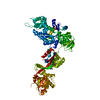
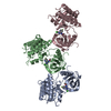
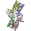
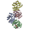
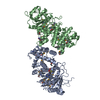
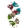
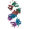
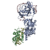
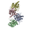
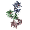
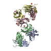
| Title | Fitting of the eEF2 crystal structure into the cryo-EM density map of the eEF2.80S.AlF4-.GDP complex | ||||||
 Components Components | Elongation factor 2 | ||||||
 Keywords Keywords | CELL CYCLE / eEF2 transition state complex / 80S Ribosome / AlF4- / GDP / GTPase / Translocation / Elongation factor / GTP-binding / Nucleotide-binding / Phosphoprotein / Protein biosynthesis / RNA-binding / rRNA-binding | ||||||
| Function / homology |  Function and homology information Function and homology informationPeptide chain elongation / Synthesis of diphthamide-EEF2 / positive regulation of translational elongation / Protein methylation / translational elongation / translation elongation factor activity / Neutrophil degranulation / maintenance of translational fidelity / Hydrolases; Acting on acid anhydrides; Acting on GTP to facilitate cellular and subcellular movement / ribosome binding ...Peptide chain elongation / Synthesis of diphthamide-EEF2 / positive regulation of translational elongation / Protein methylation / translational elongation / translation elongation factor activity / Neutrophil degranulation / maintenance of translational fidelity / Hydrolases; Acting on acid anhydrides; Acting on GTP to facilitate cellular and subcellular movement / ribosome binding / protein-folding chaperone binding / rRNA binding / ribonucleoprotein complex / GTPase activity / GTP binding / identical protein binding / cytosol Similarity search - Function | ||||||
| Biological species |  | ||||||
| Method | ELECTRON MICROSCOPY / single particle reconstruction / cryo EM / Resolution: 12.6 Å | ||||||
 Authors Authors | Sengupta, J. / Frank, J. | ||||||
 Citation Citation | Journal: J Mol Biol / Year: 2008 Title: Visualization of the eEF2-80S ribosome transition-state complex by cryo-electron microscopy. Authors: Jayati Sengupta / Jakob Nilsson / Richard Gursky / Morten Kjeldgaard / Poul Nissen / Joachim Frank /  Abstract: In an attempt to understand ribosome-induced GTP hydrolysis on eEF2, we determined a 12.6-A cryo-electron microscopy reconstruction of the eEF2-bound 80S ribosome in the presence of aluminum ...In an attempt to understand ribosome-induced GTP hydrolysis on eEF2, we determined a 12.6-A cryo-electron microscopy reconstruction of the eEF2-bound 80S ribosome in the presence of aluminum tetrafluoride and GDP, with aluminum tetrafluoride mimicking the gamma-phosphate during hydrolysis. This is the first visualization of a structure representing a transition-state complex on the ribosome. Tight interactions are observed between the factor's G domain and the large ribosomal subunit, as well as between domain IV and an intersubunit bridge. In contrast, some of the domains of eEF2 implicated in small subunit binding display a large degree of flexibility. Furthermore, we find support for a transition-state model conformation of the switch I region in this complex where the reoriented switch I region interacts with a conserved rRNA region of the 40S subunit formed by loops of the 18S RNA helices 8 and 14. This complex is structurally distinct from the eEF2-bound 80S ribosome complexes previously reported, and analysis of this map sheds light on the GTPase-coupled translocation mechanism. | ||||||
| History |
|
- Structure visualization
| Movie |
Movie viewer |
|---|---|
| Structure viewer | Molecule: MolmilJmol/JSmol |
- Downloads & links
-Download
| PDBx/mmCIF format | 3dny.cif.gz | 38.2 KB | Display | PDBx/mmCIF format |
|---|---|---|---|---|
| PDB format | pdb3dny.ent.gz | 19 KB | Display | PDB format |
| PDBx/mmJSON format | 3dny.json.gz | Tree view | PDBx/mmJSON format | |
| Others |  Other downloads Other downloads |
-Validation report
| Summary document | 3dny_validation.pdf.gz | 549.6 KB | Display | wwPDB validaton report |
|---|---|---|---|---|
| Full document | 3dny_full_validation.pdf.gz | 549.2 KB | Display | |
| Data in XML | 3dny_validation.xml.gz | 14 KB | Display | |
| Data in CIF | 3dny_validation.cif.gz | 19.7 KB | Display | |
| Arichive directory | https://data.pdbj.org/pub/pdb/validation_reports/dn/3dnyftp://data.pdbj.org/pub/pdb/validation_reports/dn/3dny | HTTPS FTP |
-Related structure data
| Related structure data | 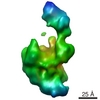 5017MC 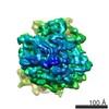 5015C 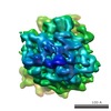 5016C 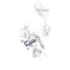 3dwuC C: citing same article ( M: map data used to model this data |
|---|---|
| Similar structure data |
-Links
 PDBj
PDBj


- Assembly
| Deposited unit | 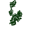
|
|---|---|
| 1 |
|
-Components
| #1: Protein | Mass: 93407.125 Da / Num. of mol.: 1 / Source method: isolated from a natural source / Source: (natural) |
|---|
-Experimental details
-Experiment
| Experiment | Method: ELECTRON MICROSCOPY |
|---|---|
| EM experiment | Aggregation state: PARTICLE / 3D reconstruction method: single particle reconstruction |
- Sample preparation
| Component | Name: eEF2-bound 80S complex in presence of AlF4-, and GDP / Type: RIBOSOME |
|---|---|
| Buffer solution | Name: 20 mM Hepes-NH3, 100 mM KCl, 20 mM MgCl2 / pH: 7.2 / Details: 20 mM Hepes-NH3, 100 mM KCl, 20 mM MgCl2 |
| Specimen | Embedding applied: NO / Shadowing applied: NO / Staining applied: NO / Vitrification applied: YES |
| Vitrification | Instrument: FEI VITROBOT MARK I / Cryogen name: ETHANE |
- Electron microscopy imaging
| Experimental equipment |  Model: Tecnai F20 / Image courtesy: FEI Company |
|---|---|
| Microscopy | Model: FEI TECNAI F20 |
| Electron gun | Electron source:  FIELD EMISSION GUN / Accelerating voltage: 200 kV / Illumination mode: FLOOD BEAM FIELD EMISSION GUN / Accelerating voltage: 200 kV / Illumination mode: FLOOD BEAM |
| Electron lens | Mode: BRIGHT FIELD / Nominal magnification: 50000 X / Calibrated magnification: 49650 X / Nominal defocus max: 1500 nm / Nominal defocus min: 4500 nm |
| Specimen holder | Temperature: 93 K |
| Image recording | Electron dose: 10 e/Å2 / Film or detector model: KODAK SO-163 FILM |
- Processing
| EM software |
| ||||||||||||
|---|---|---|---|---|---|---|---|---|---|---|---|---|---|
| CTF correction | Details: segregation in defocus groups and correction in volumes | ||||||||||||
| Symmetry | Point symmetry: C1 (asymmetric) | ||||||||||||
| 3D reconstruction | Method: Single particle 3D reconstruction / Resolution: 12.6 Å / Nominal pixel size: 2.82 Å Details: supervised classification was used (ref. Valle, M. et al, 2002, EMBO J.) Symmetry type: POINT | ||||||||||||
| Atomic model building | Protocol: RIGID BODY FIT / Space: REAL Details: METHOD--Each domain fitted as rigid body, domains I (G and G), II,IV, and V were fitted. Domain III, and domain IV insertions were not included in this fitting model. The coordinates for ...Details: METHOD--Each domain fitted as rigid body, domains I (G and G), II,IV, and V were fitted. Domain III, and domain IV insertions were not included in this fitting model. The coordinates for this entry are based on manual fitting of the coordinates of the crystal structure 1N0U into cryo-EM density map. Therefore, authors did not deposit new structure factors. | ||||||||||||
| Atomic model building | PDB-ID: 1N0U Accession code: 1N0U / Source name: PDB / Type: experimental model | ||||||||||||
| Refinement step | Cycle: LAST
|
