- PDB-2okc: Crystal structure of Type I restriction enzyme StySJI M protein (... -
+
Open data
ID or keywords:
Loading...
-
Basic information
Entry
Database: PDB / ID: 2okc
Title
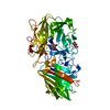
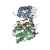
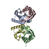
Crystal structure of Type I restriction enzyme StySJI M protein (NP_813429.1) from Bacteroides thetaiotaomicron VPI-5482 at 2.20 A resolution
Components
Type I restriction enzyme StySJI M protein
Keywords
TRANSFERASE / NP_813429.1 / N-6 DNA Methylase / Type I restriction enzyme StySJI M protein / Structural Genomics / Joint Center for Structural Genomics / JCSG / Protein Structure Initiative / PSI-2
Function / homology
Function and homology information
N-methyltransferase activity / site-specific DNA-methyltransferase (adenine-specific) / site-specific DNA-methyltransferase (adenine-specific) activity / DNA restriction-modification system / methylation / DNA binding Similarity search - Function
N6 adenine-specific DNA methyltransferase, N-terminal domain / : / N6 adenine-specific DNA methyltransferase, N-terminal domain / Type I restriction enzyme EcoKI-like, methylase subunit, N-terminal domain superfamily / HsdM N-terminal domain / N-6 DNA Methylase / DNA methylase, adenine-specific / N-6 Adenine-specific DNA methylases signature. / DNA methylase, N-6 adenine-specific, conserved site / Ferritin ...N6 adenine-specific DNA methyltransferase, N-terminal domain / : / N6 adenine-specific DNA methyltransferase, N-terminal domain / Type I restriction enzyme EcoKI-like, methylase subunit, N-terminal domain superfamily / HsdM N-terminal domain / N-6 DNA Methylase / DNA methylase, adenine-specific / N-6 Adenine-specific DNA methylases signature. / DNA methylase, N-6 adenine-specific, conserved site / Ferritin / Vaccinia Virus protein VP39 / S-adenosyl-L-methionine-dependent methyltransferase superfamily / Up-down Bundle / Rossmann fold / 3-Layer(aba) Sandwich / Mainly Alpha / Alpha Beta Similarity search - Domain/homology
ISOPROPYL ALCOHOL / S-ADENOSYLMETHIONINE / Type I restriction enzyme BthVORF4518P methylase subunit Similarity search - Component
Biological species
Bacteroides thetaiotaomicron VPI-5482 (bacteria)
Method
X-RAY DIFFRACTION / SYNCHROTRON / MAD / Resolution: 2.2 Å
BIOMOLECULE: 1, 2 THIS ENTRY CONTAINS THE CRYSTALLOGRAPHIC ASYMMETRIC UNIT WHICH CONSISTS OF 2 ... BIOMOLECULE: 1, 2 THIS ENTRY CONTAINS THE CRYSTALLOGRAPHIC ASYMMETRIC UNIT WHICH CONSISTS OF 2 CHAIN(S). SEE REMARK 350 FOR INFORMATION ON GENERATING THE BIOLOGICAL MOLECULE(S). SIZE EXCLUSION CHROMATOGRAPHY SUPPORTS THE ASSIGNMENT OF A MONOMER AS A SIGNIFICANT OLIGOMERIZATION STATE IN SOLUTION.
Remark 999
SEQUENCE THE CONSTRUCT WAS EXPRESSED WITH A PURIFICATION TAG MGSDKIHHHHHHENLYFQG. THE TAG WAS ... SEQUENCE THE CONSTRUCT WAS EXPRESSED WITH A PURIFICATION TAG MGSDKIHHHHHHENLYFQG. THE TAG WAS REMOVED WITH TEV PROTEASE LEAVING ONLY A GLYCINE FOLLOWED BY THE TARGET SEQUENCE. THE CONSTRUCT IS TRUNCATED TO EXPRESS 1-444.
Type: MARMOSAIC 325 mm CCD / Detector: CCD / Date: Dec 17, 2006 / Details: FLAT MIRROR (VERTICAL FOCUSING)
Radiation
Monochromator: SINGLE CRYSTAL SI(111) BENT (HORIZONTAL FOCUSING) Protocol: MAD / Monochromatic (M) / Laue (L): M / Scattering type: x-ray
Radiation wavelength
ID
Wavelength (Å)
Relative weight
1
0.91837
1
2
0.97899
1
3
0.97935
1
Reflection
Resolution: 2.2→28.62 Å / Num. obs: 46716 / % possible obs: 88.9 % / Redundancy: 3.97 % / Biso Wilson estimate: 45.5 Å2 / Rmerge(I) obs: 0.049 / Net I/σ(I): 11.63
Reflection shell
Resolution (Å)
Rmerge(I) obs
Mean I/σ(I) obs
Num. measured obs
Diffraction-ID
% possible all
2.2-2.28
0.378
2.2
17760
1
86.2
2.28-2.37
0.324
2.6
18322
1
90.9
2.37-2.48
0.259
3.3
18962
1
90.8
2.48-2.61
0.208
4.1
18522
1
90.3
2.61-2.77
0.147
6
18283
1
90.3
2.77-2.98
0.101
8.4
18433
1
90
2.98-3.28
0.064
12.6
18829
1
88.9
3.28-3.76
0.04
19.8
18921
1
88.7
3.76-4.72
0.027
26.8
18603
1
88.1
4.72-28.6
0.021
31.3
18610
1
85.3
-
Phasing
Phasing
Method: MAD
-
Processing
Software
Name
Version
Classification
NB
MolProbity
3beta29
modelbuilding
SOLVE
phasing
REFMAC
5.2.0005
refinement
XSCALE
datascaling
PDB_EXTRACT
2
dataextraction
XDS
datareduction
Refinement
Method to determine structure: MAD / Resolution: 2.2→28.62 Å / Cor.coef. Fo:Fc: 0.959 / Cor.coef. Fo:Fc free: 0.942 / SU B: 12.486 / SU ML: 0.165 / TLS residual ADP flag: LIKELY RESIDUAL / Cross valid method: THROUGHOUT / σ(F): 0 / ESU R: 0.28 / ESU R Free: 0.214 Stereochemistry target values: MAXIMUM LIKELIHOOD WITH PHASES Details: 1. HYDROGENS HAVE BEEN ADDED IN THE RIDING POSITIONS. 2. A MET-INHIBITION PROTOCOL WAS USED FOR SELENOMETHIONINE INCORPORATION DURING PROTEIN EXPRESSION. THE OCCUPANCY OF THE SE ATOMS IN THE ...Details: 1. HYDROGENS HAVE BEEN ADDED IN THE RIDING POSITIONS. 2. A MET-INHIBITION PROTOCOL WAS USED FOR SELENOMETHIONINE INCORPORATION DURING PROTEIN EXPRESSION. THE OCCUPANCY OF THE SE ATOMS IN THE MSE RESIDUES WAS REDUCED TO 0.75 FOR THE REDUCED SCATTERING POWER DUE TO PARTIAL S-MET INCORPORATION. 3. ATOM RECORD CONTAINS RESIDUAL B FACTORS ONLY. 4. S-ADENOSYLMETHIONINES WERE MODELED BASED ON PROPOSED FUNCTION AND STRUCTURAL HOMOLOGS. 5. CHLORINE, GLYCEROL AND 2- PROPANOL WERE MODELED BASED ON CRYSTALLIZATION CONDITIONS. 6. RAMACHANDRAN OUTLIERS A124 AND B9 HAVE WEAK ELECTRON DENSITY. 7. RESIDUES A1-8, A197, A434-444, B1-6, AND B433-444 ARE IN DISORDERED REGIONS AND WERE NOT MODELED.
Rfactor
Num. reflection
% reflection
Selection details
Rfree
0.232
2367
5.1 %
RANDOM
Rwork
0.182
-
-
-
obs
0.185
46612
91.8 %
-
Solvent computation
Ion probe radii: 0.8 Å / Shrinkage radii: 0.8 Å / VDW probe radii: 1.2 Å / Solvent model: MASK
In the structure databanks used in Yorodumi, some data are registered as the other names, "COVID-19 virus" and "2019-nCoV". Here are the details of the virus and the list of structure data.
Jan 31, 2019. EMDB accession codes are about to change! (news from PDBe EMDB page)
EMDB accession codes are about to change! (news from PDBe EMDB page)
The allocation of 4 digits for EMDB accession codes will soon come to an end. Whilst these codes will remain in use, new EMDB accession codes will include an additional digit and will expand incrementally as the available range of codes is exhausted. The current 4-digit format prefixed with “EMD-” (i.e. EMD-XXXX) will advance to a 5-digit format (i.e. EMD-XXXXX), and so on. It is currently estimated that the 4-digit codes will be depleted around Spring 2019, at which point the 5-digit format will come into force.
The EM Navigator/Yorodumi systems omit the EMD- prefix.
Related info.:Q: What is EMD? / ID/Accession-code notation in Yorodumi/EM Navigator
Yorodumi is a browser for structure data from EMDB, PDB, SASBDB, etc.
This page is also the successor to EM Navigator detail page, and also detail information page/front-end page for Omokage search.
The word "yorodu" (or yorozu) is an old Japanese word meaning "ten thousand". "mi" (miru) is to see.
Related info.:EMDB / PDB / SASBDB / Comparison of 3 databanks / Yorodumi Search / Aug 31, 2016. New EM Navigator & Yorodumi / Yorodumi Papers / Jmol/JSmol / Function and homology information / Changes in new EM Navigator and Yorodumi
 Movie
Movie Controller

 Yorodumi
 Open data
 Basic information
 Components
 Keywords
 Function and homology information
 Bacteroides thetaiotaomicron VPI-5482 (bacteria)
 X-RAY DIFFRACTION /
  Authors
 Citation
 Structure visualization
 Downloads & links
 Other downloads
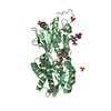


 PDBj
PDBj

 Assembly
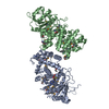


 Mass: 398.437 Da / Num. of mol.: 2 / Source method: obtained synthetically / Formula: C15H22N6O5S
 Mass: 92.094 Da / Num. of mol.: 1 / Source method: obtained synthetically / Formula: C3H8O3
 Mass: 60.095 Da / Num. of mol.: 2 / Source method: obtained synthetically / Formula: C3H8O
 Mass: 35.453 Da / Num. of mol.: 1 / Source method: obtained synthetically / Formula: Cl
 Sample preparation
 / Beamline: BL11-1 / Wavelength: 0.91837, 0.97899, 0.97935
 Processing