 Movie
Movie Controller

[English] 日本語
 Yorodumi
- PDB-3dwu: Transition-state model conformation of the switch I region fitted... -
+ Open data

- Basic information
| Entry | Database: PDB / ID: 3dwu | ||||||
|---|---|---|---|---|---|---|---|
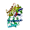
| Title | Transition-state model conformation of the switch I region fitted into the cryo-EM map of the eEF2.80S.AlF4.GDP complex | ||||||
 Components Components | Elongation factor Tu-B | ||||||
 Keywords Keywords | BIOSYNTHETIC PROTEIN / Transition state / conserved switch I / Antibiotic resistance / Elongation factor / GTP-binding / Membrane / Methylation / Nucleotide-binding / Phosphoprotein / Protein biosynthesis | ||||||
| Function / homology |  Function and homology information Function and homology informationprotein-synthesizing GTPase / translation elongation factor activity / GTPase activity / GTP binding / magnesium ion binding / cytosol Similarity search - Function | ||||||
| Biological species |   Thermus thermophilus HB8 (bacteria) Thermus thermophilus HB8 (bacteria) | ||||||
| Method | ELECTRON MICROSCOPY / single particle reconstruction / cryo EM / Resolution: 12.6 Å | ||||||
 Authors Authors | Nissen, P. / Nyborg, J. / Kjeldgaard, M. | ||||||
 Citation Citation | Journal: J Mol Biol / Year: 2008 Title: Visualization of the eEF2-80S ribosome transition-state complex by cryo-electron microscopy. Authors: Jayati Sengupta / Jakob Nilsson / Richard Gursky / Morten Kjeldgaard / Poul Nissen / Joachim Frank /  Abstract: In an attempt to understand ribosome-induced GTP hydrolysis on eEF2, we determined a 12.6-A cryo-electron microscopy reconstruction of the eEF2-bound 80S ribosome in the presence of aluminum ...In an attempt to understand ribosome-induced GTP hydrolysis on eEF2, we determined a 12.6-A cryo-electron microscopy reconstruction of the eEF2-bound 80S ribosome in the presence of aluminum tetrafluoride and GDP, with aluminum tetrafluoride mimicking the gamma-phosphate during hydrolysis. This is the first visualization of a structure representing a transition-state complex on the ribosome. Tight interactions are observed between the factor's G domain and the large ribosomal subunit, as well as between domain IV and an intersubunit bridge. In contrast, some of the domains of eEF2 implicated in small subunit binding display a large degree of flexibility. Furthermore, we find support for a transition-state model conformation of the switch I region in this complex where the reoriented switch I region interacts with a conserved rRNA region of the 40S subunit formed by loops of the 18S RNA helices 8 and 14. This complex is structurally distinct from the eEF2-bound 80S ribosome complexes previously reported, and analysis of this map sheds light on the GTPase-coupled translocation mechanism. | ||||||
| History |
|
- Structure visualization
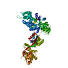
| Movie |
Movie viewer |
|---|---|
| Structure viewer | Molecule: MolmilJmol/JSmol |
- Downloads & links
-Download
| PDBx/mmCIF format | 3dwu.cif.gz | 10.7 KB | Display | PDBx/mmCIF format |
|---|---|---|---|---|
| PDB format | pdb3dwu.ent.gz | 3.8 KB | Display | PDB format |
| PDBx/mmJSON format | 3dwu.json.gz | Tree view | PDBx/mmJSON format | |
| Others |  Other downloads Other downloads |
-Validation report
| Arichive directory | https://data.pdbj.org/pub/pdb/validation_reports/dw/3dwuftp://data.pdbj.org/pub/pdb/validation_reports/dw/3dwu | HTTPS FTP |
|---|
-Related structure data
| Related structure data | 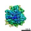 5015MC 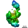 5017MC 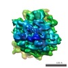 5016C 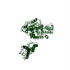 3dnyC C: citing same article ( M: map data used to model this data |
|---|---|
| Similar structure data |
-Links
 PDBj
PDBj


- Assembly
| Deposited unit | 
|
|---|---|
| 1 |
|
-Components
| #1: Protein/peptide | Mass: 4949.441 Da / Num. of mol.: 1 / Fragment: Switch I region: UNP residues 21-66 / Source method: isolated from a natural source / Source: (natural) Thermus thermophilus HB8 (bacteria) / Strain: HB8 / DSM 579 / References: UniProt: P60339 |
|---|
-Experimental details
-Experiment
| Experiment | Method: ELECTRON MICROSCOPY |
|---|---|
| EM experiment | Aggregation state: PARTICLE / 3D reconstruction method: single particle reconstruction |
- Sample preparation
| Component | Name: eEF2.80S.AlF4.GDP complex / Type: RIBOSOME |
|---|---|
| Buffer solution | Name: 20 mM Hepes-NH3, 100 mM KCl, 20 mM MgCl2 / pH: 7.2 / Details: 20 mM Hepes-NH3, 100 mM KCl, 20 mM MgCl2 |
| Specimen | Embedding applied: NO / Shadowing applied: NO / Staining applied: NO / Vitrification applied: YES |
| Vitrification | Instrument: FEI VITROBOT MARK I / Cryogen name: NITROGEN Details: Cryogen ETHANE (93K), two-face blotting for 1 second |
- Electron microscopy imaging
| Experimental equipment |  Model: Tecnai F20 / Image courtesy: FEI Company |
|---|---|
| Microscopy | Model: FEI TECNAI F20 |
| Electron gun | Electron source:  FIELD EMISSION GUN / Accelerating voltage: 200 kV / Illumination mode: FLOOD BEAM FIELD EMISSION GUN / Accelerating voltage: 200 kV / Illumination mode: FLOOD BEAM |
| Electron lens | Mode: BRIGHT FIELD / Nominal magnification: 50000 X / Calibrated magnification: 49650 X / Nominal defocus max: 4500 nm / Nominal defocus min: 1500 nm |
| Image recording | Electron dose: 10 e/Å2 / Details: Kodak SO163 film |
- Processing
| EM software |
| ||||||||||||
|---|---|---|---|---|---|---|---|---|---|---|---|---|---|
| CTF correction | Details: segregation in defocus groups and correction in volumes | ||||||||||||
| Symmetry | Point symmetry: C1 (asymmetric) | ||||||||||||
| 3D reconstruction | Method: SPIDER / Resolution: 12.6 Å / Resolution method: FSC / Num. of particles: 28242 / Nominal pixel size: 2.82 Å Details: Single particle reconstruction, resolution estimated: FSC cut-off at 0.15 Symmetry type: POINT | ||||||||||||
| Atomic model building | Protocol: RIGID BODY FIT / Space: REAL / Target criteria: correlation coefficient Details: METHOD--manual REFINEMENT PROTOCOL--Fitted as rigid body. Current model was aligned to the helix A of the fitted eEF2 coordinates (PDB entry 3DNY) which is located next to the switch I ...Details: METHOD--manual REFINEMENT PROTOCOL--Fitted as rigid body. Current model was aligned to the helix A of the fitted eEF2 coordinates (PDB entry 3DNY) which is located next to the switch I sequence. The coordinates for this entry are based on manual fitting of the coordinates into cryo-EM density map. Therefore, authors did not deposit structure factors. | ||||||||||||
| Atomic model building | PDB-ID: 3DNY Accession code: 3DNY / Source name: PDB / Type: experimental model | ||||||||||||
| Refinement step | Cycle: LAST /
|
