 Movie
Movie Controller

[English] 日本語
 Yorodumi
- PDB-6c31: Crystal structure of TetR family protein Rv0078 in complex with DNA -
+ Open data

- Basic information
| Entry | Database: PDB / ID: 6c31 | ||||||
|---|---|---|---|---|---|---|---|
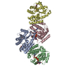
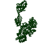
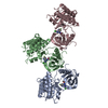
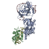
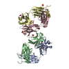
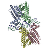
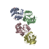
| Title | Crystal structure of TetR family protein Rv0078 in complex with DNA | ||||||
 Components Components |
| ||||||
 Keywords Keywords | DNA BINDING PROTEIN/DNA / Rv0078 / DNA / TetR family / DNA BINDING PROTEIN-DNA complex | ||||||
| Function / homology |  Function and homology information Function and homology informationtranscription cis-regulatory region binding / DNA-binding transcription factor activity / regulation of DNA-templated transcription / plasma membrane Similarity search - Function | ||||||
| Biological species |   Mycobacterium tuberculosis (bacteria)Mycobacterium tuberculosis H37Rv (bacteria) Mycobacterium tuberculosis (bacteria)Mycobacterium tuberculosis H37Rv (bacteria) | ||||||
| Method |  X-RAY DIFFRACTION / SYNCHROTRON / MOLECULAR REPLACEMENT / Resolution: 3 Å X-RAY DIFFRACTION / SYNCHROTRON / MOLECULAR REPLACEMENT / Resolution: 3 Å | ||||||
 Authors Authors | Hsu, H.C. / Li, H. | ||||||
| Funding support |  United States, 1items United States, 1items
| ||||||
 Citation Citation | Journal: MBio / Year: 2018 Title: Cytokinin Signaling in Mycobacterium tuberculosis. Authors: Samanovic, M.I. / Hsu, H.C. / Jones, M.B. / Jones, V. / McNeil, M.R. / Becker, S.H. / Jordan, A.T. / Strnad, M. / Xu, C. / Jackson, M. / Li, H. / Darwin, K.H. | ||||||
| History |
|
- Structure visualization
| Structure viewer | Molecule: MolmilJmol/JSmol |
|---|
- Downloads & links
-Download
| PDBx/mmCIF format | 6c31.cif.gz | 346.3 KB | Display | PDBx/mmCIF format |
|---|---|---|---|---|
| PDB format | pdb6c31.ent.gz | 275.5 KB | Display | PDB format |
| PDBx/mmJSON format | 6c31.json.gz | Tree view | PDBx/mmJSON format | |
| Others |  Other downloads Other downloads |
-Validation report
| Arichive directory | https://data.pdbj.org/pub/pdb/validation_reports/c3/6c31ftp://data.pdbj.org/pub/pdb/validation_reports/c3/6c31 | HTTPS FTP |
|---|
-Related structure data
| Related structure data | 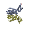 5wm9SC S: Starting model for refinement C: citing same article ( |
|---|---|
| Similar structure data |


-Links
 PDBj
PDBj


- Assembly
| Deposited unit | 
| ||||||||||||
|---|---|---|---|---|---|---|---|---|---|---|---|---|---|
| 1 | 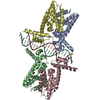
| ||||||||||||
| 2 | 
| ||||||||||||
| Unit cell |
|
-Components
| #1: Protein | Mass: 23477.922 Da / Num. of mol.: 8 Source method: isolated from a genetically manipulated source Source: (gene. exp.) Mycobacterium tuberculosis (strain ATCC 25618 / H37Rv) (bacteria)Strain: ATCC 25618 / H37Rv / Gene: LH57_00450 / Production host: #2: DNA chain | Mass: 7049.574 Da / Num. of mol.: 2 / Source method: obtained synthetically Source: (synth.) Mycobacterium tuberculosis H37Rv (bacteria)#3: DNA chain | Mass: 7071.570 Da / Num. of mol.: 2 / Source method: obtained synthetically Source: (synth.) Mycobacterium tuberculosis H37Rv (bacteria) |
|---|
-Experimental details
-Experiment
| Experiment | Method: X-RAY DIFFRACTION / Number of used crystals: 1 |
|---|
- Sample preparation
| Crystal | Density Matthews: 2.84 Å3/Da / Density % sol: 56.63 % |
|---|---|
| Crystal grow | Temperature: 293 K / Method: vapor diffusion, sitting drop / Details: 0.2 M magnesium formate dihydrate |
-Data collection
| Diffraction | Mean temperature: 100 K |
|---|---|
| Diffraction source | Source: SYNCHROTRON / Site: APS / Beamline: 21-ID-D / Wavelength: 1.12713 Å |
| Detector | Type: MARMOSAIC 300 mm CCD / Detector: CCD / Date: Aug 7, 2017 |
| Radiation | Monochromator: Si(111) / Protocol: SINGLE WAVELENGTH / Monochromatic (M) / Laue (L): M / Scattering type: x-ray |
| Radiation wavelength | Wavelength: 1.12713 Å / Relative weight: 1 |
| Reflection | Resolution: 3→28.74 Å / Num. obs: 46330 / % possible obs: 94.8 % / Redundancy: 6.4 % / Rpim(I) all: 0.087 / Net I/σ(I): 5.5 |
| Reflection shell | Resolution: 3→3.16 Å / Rpim(I) all: 0.474 |
- Processing
| Software |
| ||||||||||||||||||||
|---|---|---|---|---|---|---|---|---|---|---|---|---|---|---|---|---|---|---|---|---|---|
| Refinement | Method to determine structure: MOLECULAR REPLACEMENT Starting model: PDB entry 5WM9 Resolution: 3→28.495 Å / Cross valid method: FREE R-VALUE
| ||||||||||||||||||||
| Displacement parameters | Biso mean: 73 Å2 | ||||||||||||||||||||
| Refinement step | Cycle: LAST / Resolution: 3→28.495 Å
|
