 Movie
Movie Controller

[English] 日本語
 Yorodumi
- PDB-3znp: IN VITRO AND IN VIVO INHIBITION OF HUMAN D-AMINO ACID OXIDASE: RE... -
+ Open data

- Basic information
| Entry | Database: PDB / ID: 3znp | ||||||
|---|---|---|---|---|---|---|---|
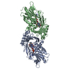
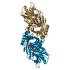
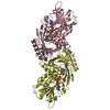
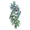
| Title | IN VITRO AND IN VIVO INHIBITION OF HUMAN D-AMINO ACID OXIDASE: REGULATION OF D-SERINE CONCENTRATION IN THE BRAIN | ||||||
 Components Components | D-AMINO-ACID OXIDASE | ||||||
 Keywords Keywords | OXIDOREDUCTASE / SMALL MOLECULE INHIBITION / NEUROTRANSMISSION | ||||||
| Function / homology |  Function and homology information Function and homology informationD-amino-acid dehydrogenase activity / D-amino-acid oxidase / D-amino-acid oxidase activity / D-alanine catabolic process / : / glycine oxidase activity / L-proline catabolic process / D-amino acid catabolic process / D-serine catabolic process / Glyoxylate metabolism and glycine degradation ...D-amino-acid dehydrogenase activity / D-amino-acid oxidase / D-amino-acid oxidase activity / D-alanine catabolic process / : / glycine oxidase activity / L-proline catabolic process / D-amino acid catabolic process / D-serine catabolic process / Glyoxylate metabolism and glycine degradation / presynaptic active zone / neutrophil-mediated killing of gram-negative bacterium / dopamine biosynthetic process / peroxisomal matrix / digestion / FAD binding / Peroxisomal protein import / : / identical protein binding / cytosol / cytoplasm Similarity search - Function | ||||||
| Biological species |  HOMO SAPIENS (human) HOMO SAPIENS (human) | ||||||
| Method |  X-RAY DIFFRACTION / SYNCHROTRON / MOLECULAR REPLACEMENT / Resolution: 2.4 Å X-RAY DIFFRACTION / SYNCHROTRON / MOLECULAR REPLACEMENT / Resolution: 2.4 Å | ||||||
 Authors Authors | Hopkins, S.C. / Heffernan, M.L.R. / Saraswat, L.D. / Bowen, C.A. / Melnick, L. / Hardy, L.W. / Orsini, M.A. / Allen, M.S. / Koch, P. / Spear, K.L. ...Hopkins, S.C. / Heffernan, M.L.R. / Saraswat, L.D. / Bowen, C.A. / Melnick, L. / Hardy, L.W. / Orsini, M.A. / Allen, M.S. / Koch, P. / Spear, K.L. / Foglesong, R.J. / Soukri, M. / Chytil, M. / Fang, Q.K. / Jones, S.W. / Varney, M.A. / Panatier, A. / Oliet, S.H.R. / Pollegioni, L. / Piubelli, L. / Molla, G. / Nardini, M. / Large, T.H. | ||||||
 Citation Citation | Journal: J.Med.Chem. / Year: 2013 Title: Structural, Kinetic, and Pharmacodynamic Mechanisms of D-Amino Acid Oxidase Inhibition by Small Molecules. Authors: Hopkins, S.C. / Heffernan, M.L.R. / Saraswat, L.D. / Bowen, C.A. / Melnick, L. / Hardy, L.W. / Orsini, M.A. / Allen, M.S. / Koch, P. / Spear, K.L. / Foglesong, R.J. / Soukri, M. / Chytil, M. ...Authors: Hopkins, S.C. / Heffernan, M.L.R. / Saraswat, L.D. / Bowen, C.A. / Melnick, L. / Hardy, L.W. / Orsini, M.A. / Allen, M.S. / Koch, P. / Spear, K.L. / Foglesong, R.J. / Soukri, M. / Chytil, M. / Fang, Q.K. / Jones, S.W. / Varney, M.A. / Panatier, A. / Oliet, S.H.R. / Pollegioni, L. / Piubelli, L. / Molla, G. / Nardini, M. / Large, T.H. | ||||||
| History |
|
- Structure visualization
| Structure viewer | Molecule: MolmilJmol/JSmol |
|---|
- Downloads & links
-Download
| PDBx/mmCIF format | 3znp.cif.gz | 305.4 KB | Display | PDBx/mmCIF format |
|---|---|---|---|---|
| PDB format | pdb3znp.ent.gz | 250.2 KB | Display | PDB format |
| PDBx/mmJSON format | 3znp.json.gz | Tree view | PDBx/mmJSON format | |
| Others |  Other downloads Other downloads |
-Validation report
| Arichive directory | https://data.pdbj.org/pub/pdb/validation_reports/zn/3znpftp://data.pdbj.org/pub/pdb/validation_reports/zn/3znp | HTTPS FTP |
|---|
-Related structure data
| Related structure data |  3znnC  3znoC  3znqC  2du8S C: citing same article ( S: Starting model for refinement |
|---|---|
| Similar structure data |


-Links
 PDBj
PDBj- Assembly
| Deposited unit | 
| ||||||||
|---|---|---|---|---|---|---|---|---|---|
| 1 | 
| ||||||||
| 2 | 
| ||||||||
| Unit cell |
|
-Components
| #1: Protein | Mass: 39520.910 Da / Num. of mol.: 2 Source method: isolated from a genetically manipulated source Source: (gene. exp.) HOMO SAPIENS (human) / Production host:  #2: Chemical |   Mass: 785.550 Da / Num. of mol.: 2 / Source method: obtained synthetically / Formula: C27H33N9O15P2 / Comment: FAD*YM Mass: 785.550 Da / Num. of mol.: 2 / Source method: obtained synthetically / Formula: C27H33N9O15P2 / Comment: FAD*YM#3: Chemical | ChemComp-SE2 / |   Mass: 162.142 Da / Num. of mol.: 1 / Source method: obtained synthetically / Formula: C9H6O3 Mass: 162.142 Da / Num. of mol.: 1 / Source method: obtained synthetically / Formula: C9H6O3#4: Chemical | ChemComp-GOL /   Mass: 92.094 Da / Num. of mol.: 7 / Source method: obtained synthetically / Formula: C3H8O3 Mass: 92.094 Da / Num. of mol.: 7 / Source method: obtained synthetically / Formula: C3H8O3#5: Water | ChemComp-HOH / |  Mass: 18.015 Da / Num. of mol.: 99 / Source method: isolated from a natural source / Formula: H2O Mass: 18.015 Da / Num. of mol.: 99 / Source method: isolated from a natural source / Formula: H2O |
|---|
-Experimental details
-Experiment
| Experiment | Method: X-RAY DIFFRACTION / Number of used crystals: 1 |
|---|
- Sample preparation
| Crystal | Density Matthews: 2.44 Å3/Da / Density % sol: 49.62 % / Description: NONE |
|---|
-Data collection
| Diffraction | Mean temperature: 100 K |
|---|---|
| Diffraction source | Source: SYNCHROTRON / Site: ALS  / Type: ALS / Wavelength: 1.24 / Type: ALS / Wavelength: 1.24 |
| Radiation | Protocol: SINGLE WAVELENGTH / Monochromatic (M) / Laue (L): M / Scattering type: x-ray |
| Radiation wavelength | Wavelength: 1.24 Å / Relative weight: 1 |
| Reflection | Resolution: 2.4→47.66 Å / Num. obs: 30588 / % possible obs: 98.5 % / Observed criterion σ(I): 2 / Redundancy: 3.27 % / Biso Wilson estimate: 67.89 Å2 / Rmerge(I) obs: 0.08 / Net I/σ(I): 7.7 |
| Reflection shell | Resolution: 2.4→2.49 Å / Redundancy: 3.27 % / Rmerge(I) obs: 0.46 / Mean I/σ(I) obs: 1.9 / % possible all: 99.5 |
- Processing
| Software |
| ||||||||||||||||||||||||||||||||||||||||||||||||||||||||||||||||||||||||||||||||||||||||||||||||||||||||||||||||||
|---|---|---|---|---|---|---|---|---|---|---|---|---|---|---|---|---|---|---|---|---|---|---|---|---|---|---|---|---|---|---|---|---|---|---|---|---|---|---|---|---|---|---|---|---|---|---|---|---|---|---|---|---|---|---|---|---|---|---|---|---|---|---|---|---|---|---|---|---|---|---|---|---|---|---|---|---|---|---|---|---|---|---|---|---|---|---|---|---|---|---|---|---|---|---|---|---|---|---|---|---|---|---|---|---|---|---|---|---|---|---|---|---|---|---|---|
| Refinement | Method to determine structure: MOLECULAR REPLACEMENT Starting model: PDB ENTRY 2DU8 Resolution: 2.4→34.98 Å / Cor.coef. Fo:Fc: 0.9329 / Cor.coef. Fo:Fc free: 0.8818 / SU R Cruickshank DPI: 0.5 / Cross valid method: THROUGHOUT / σ(F): 0 / SU R Blow DPI: 0.503 / SU Rfree Blow DPI: 0.318 / SU Rfree Cruickshank DPI: 0.323 Details: DISORDERED REGIONS WERE MODELED STEREOCHEMICALLY. POOR DENSITY IS PRESENT FOR RESIDUES A28, A29, A30, A31, A297, A298, A299, A300, A301, A302, A303, A335, A336, A337, A338, A339, A340, B24, ...Details: DISORDERED REGIONS WERE MODELED STEREOCHEMICALLY. POOR DENSITY IS PRESENT FOR RESIDUES A28, A29, A30, A31, A297, A298, A299, A300, A301, A302, A303, A335, A336, A337, A338, A339, A340, B24, B25, B26, B27, B28, B29, B30, B31, B32, B55, B56, B57, B58, B59, B60, B61, B62, B85, B117, B123, B128, B129, B166, B167, B168, B188, B189, B190, B191, B192, B193, B194, B195, B196, B220, B221, B222, B223, B224, B253, B254, B255, B256, B260, B261, B295, B296, B297, B298, B299, B300, B301, B302, B303, B335, B336, B337, B338, B339, B340. FINAL STRUCTURE HAS NO RESIDUES IN THE DISALLOWED REGION OF THE RAMACHANDRAN PLOT AS DEFINED IN THE CCP4 PROCHECK PROGRAM.
| ||||||||||||||||||||||||||||||||||||||||||||||||||||||||||||||||||||||||||||||||||||||||||||||||||||||||||||||||||
| Displacement parameters | Biso mean: 88.17 Å2
| ||||||||||||||||||||||||||||||||||||||||||||||||||||||||||||||||||||||||||||||||||||||||||||||||||||||||||||||||||
| Refine analyze | Luzzati coordinate error obs: 0.582 Å | ||||||||||||||||||||||||||||||||||||||||||||||||||||||||||||||||||||||||||||||||||||||||||||||||||||||||||||||||||
| Refinement step | Cycle: LAST / Resolution: 2.4→34.98 Å
| ||||||||||||||||||||||||||||||||||||||||||||||||||||||||||||||||||||||||||||||||||||||||||||||||||||||||||||||||||
| Refine LS restraints |
| ||||||||||||||||||||||||||||||||||||||||||||||||||||||||||||||||||||||||||||||||||||||||||||||||||||||||||||||||||
| LS refinement shell | Resolution: 2.4→2.48 Å / Total num. of bins used: 15
| ||||||||||||||||||||||||||||||||||||||||||||||||||||||||||||||||||||||||||||||||||||||||||||||||||||||||||||||||||
| Refinement TLS params. | Method: refined / Refine-ID: X-RAY DIFFRACTION
| ||||||||||||||||||||||||||||||||||||||||||||||||||||||||||||||||||||||||||||||||||||||||||||||||||||||||||||||||||
| Refinement TLS group |
|
