 Movie
Movie Controller


+ Open data

- Basic information
| Entry | Database: PDB / ID: 1an9 | |||||||||
|---|---|---|---|---|---|---|---|---|---|---|
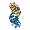
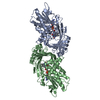
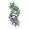
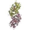
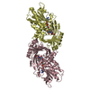
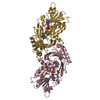
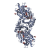
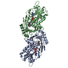
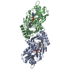
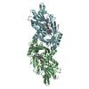
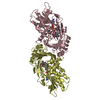
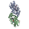
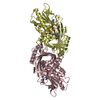
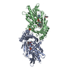
| Title | D-AMINO ACID OXIDASE COMPLEX WITH O-AMINOBENZOATE | |||||||||
 Components Components | D-AMINO ACID OXIDASE | |||||||||
 Keywords Keywords | OXIDOREDUCTASE / FAD / OXIDASE / D-AMINO ACID / FLAVOPROTEIN | |||||||||
| Function / homology |  Function and homology information Function and homology informationGlyoxylate metabolism and glycine degradation / Peroxisomal protein import / D-amino-acid oxidase / D-amino-acid oxidase activity / D-alanine catabolic process / glycine oxidase activity / L-proline catabolic process / D-amino acid catabolic process / D-serine catabolic process / presynaptic active zone ...Glyoxylate metabolism and glycine degradation / Peroxisomal protein import / D-amino-acid oxidase / D-amino-acid oxidase activity / D-alanine catabolic process / glycine oxidase activity / L-proline catabolic process / D-amino acid catabolic process / D-serine catabolic process / presynaptic active zone / neutrophil-mediated killing of gram-negative bacterium / dopamine biosynthetic process / peroxisomal matrix / digestion / FAD binding / peroxisome / extracellular region / cytosol / cytoplasm Similarity search - Function | |||||||||
| Biological species |  | |||||||||
| Method |  X-RAY DIFFRACTION / SYNCHROTRON / molecular replacement / Resolution: 2.5 Å X-RAY DIFFRACTION / SYNCHROTRON / molecular replacement / Resolution: 2.5 Å | |||||||||
 Authors Authors | Miura, R. / Setoyama, C. / Nishina, Y. / Shiga, K. / Mizutani, H. / Miyahara, I. / Hirotsu, K. | |||||||||
 Citation Citation | Journal: J.Biochem.(Tokyo) / Year: 1997 Title: Structural and mechanistic studies on D-amino acid oxidase x substrate complex: implications of the crystal structure of enzyme x substrate analog complex. Authors: Miura, R. / Setoyama, C. / Nishina, Y. / Shiga, K. / Mizutani, H. / Miyahara, I. / Hirotsu, K. #1: Journal: J.Biochem.(Tokyo) / Year: 1996Title: Three-Dimensional Structure of Porcine Kidney D-Amino Acid Oxidase at 3.0 A Resolution Authors: Mizutani, H. / Miyahara, I. / Hirotsu, K. / Nishina, Y. / Shiga, K. / Setoyama, C. / Miura, R. | |||||||||
| History |
|
- Structure visualization
| Structure viewer | Molecule: MolmilJmol/JSmol |
|---|
- Downloads & links
-Download
| PDBx/mmCIF format | 1an9.cif.gz | 150.5 KB | Display | PDBx/mmCIF format |
|---|---|---|---|---|
| PDB format | pdb1an9.ent.gz | 118.8 KB | Display | PDB format |
| PDBx/mmJSON format | 1an9.json.gz | Tree view | PDBx/mmJSON format | |
| Others |  Other downloads Other downloads |
-Validation report
| Arichive directory | https://data.pdbj.org/pub/pdb/validation_reports/an/1an9ftp://data.pdbj.org/pub/pdb/validation_reports/an/1an9 | HTTPS FTP |
|---|
-Related structure data
| Related structure data |  1aa8 S: Starting model for refinement |
|---|---|
| Similar structure data |


-Links
 PDBj
PDBj- Assembly
| Deposited unit | 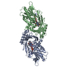
| ||||||||
|---|---|---|---|---|---|---|---|---|---|
| 1 |
| ||||||||
| Unit cell |
|
-Components
| #1: Protein | Mass: 38612.906 Da / Num. of mol.: 2 Source method: isolated from a genetically manipulated source Source: (gene. exp.)  #2: Chemical |   Mass: 785.550 Da / Num. of mol.: 2 / Source method: obtained synthetically / Formula: C27H33N9O15P2 / Comment: FAD*YM Mass: 785.550 Da / Num. of mol.: 2 / Source method: obtained synthetically / Formula: C27H33N9O15P2 / Comment: FAD*YM#3: Chemical |   Type: L-peptide linking / Mass: 137.136 Da / Num. of mol.: 2 / Source method: obtained synthetically / Formula: C7H7NO2 Type: L-peptide linking / Mass: 137.136 Da / Num. of mol.: 2 / Source method: obtained synthetically / Formula: C7H7NO2#4: Water | ChemComp-HOH / |  Mass: 18.015 Da / Num. of mol.: 112 / Source method: isolated from a natural source / Formula: H2O Mass: 18.015 Da / Num. of mol.: 112 / Source method: isolated from a natural source / Formula: H2O |
|---|
-Experimental details
-Experiment
| Experiment | Method: X-RAY DIFFRACTION / Number of used crystals: 1 |
|---|
- Sample preparation
| Crystal | Density Matthews: 2.3 Å3/Da / Density % sol: 46 % | |||||||||||||||||||||||||||||||||||||||||||||
|---|---|---|---|---|---|---|---|---|---|---|---|---|---|---|---|---|---|---|---|---|---|---|---|---|---|---|---|---|---|---|---|---|---|---|---|---|---|---|---|---|---|---|---|---|---|---|
| Crystal grow | pH: 6.3 Details: PROTEIN WAS CRYSTALLIZED FROM 120MM SODIUM ACETATE, 60MM SODIUM CITRATE, 30% PEG4000, pH 6.3 | |||||||||||||||||||||||||||||||||||||||||||||
| Crystal grow | *PLUS Temperature: 20 ℃ / Method: vapor diffusion / Details: Setoyama, C., (1996) J. Biochem., 119, 1114. | |||||||||||||||||||||||||||||||||||||||||||||
| Components of the solutions | *PLUS
|
-Data collection
| Diffraction | Mean temperature: 287 K |
|---|---|
| Diffraction source | Source: SYNCHROTRON / Site: Photon Factory  / Beamline: BL-6A / Wavelength: 1 / Beamline: BL-6A / Wavelength: 1 |
| Detector | Type: FUJI / Detector: IMAGE PLATE / Date: Nov 1, 1996 / Details: MIRROR |
| Radiation | Monochromatic (M) / Laue (L): M / Scattering type: x-ray |
| Radiation wavelength | Wavelength: 1 Å / Relative weight: 1 |
| Reflection | Resolution: 2.5→20 Å / Num. obs: 22424 / % possible obs: 87.5 % / Observed criterion σ(I): 1 / Rmerge(I) obs: 0.058 |
| Reflection shell | Resolution: 2.5→2.61 Å / % possible all: 73.32 |
| Reflection | *PLUS Num. measured all: 66408 |
- Processing
| Software |
| ||||||||||||||||||||||||||||||||||||||||||||||||||||||||||||
|---|---|---|---|---|---|---|---|---|---|---|---|---|---|---|---|---|---|---|---|---|---|---|---|---|---|---|---|---|---|---|---|---|---|---|---|---|---|---|---|---|---|---|---|---|---|---|---|---|---|---|---|---|---|---|---|---|---|---|---|---|---|
| Refinement | Method to determine structure: molecular replacement Starting model: PDB ENTRY 1AA8 1aa8 Resolution: 2.5→10 Å / Data cutoff high absF: 100000000000 / Data cutoff low absF: 0 / Cross valid method: THROUGHOUT / σ(F): 2
| ||||||||||||||||||||||||||||||||||||||||||||||||||||||||||||
| Refine analyze | Luzzati coordinate error obs: 0.28 Å | ||||||||||||||||||||||||||||||||||||||||||||||||||||||||||||
| Refinement step | Cycle: LAST / Resolution: 2.5→10 Å
| ||||||||||||||||||||||||||||||||||||||||||||||||||||||||||||
| Refine LS restraints |
| ||||||||||||||||||||||||||||||||||||||||||||||||||||||||||||
| LS refinement shell | Resolution: 2.5→2.61 Å / Total num. of bins used: 8
| ||||||||||||||||||||||||||||||||||||||||||||||||||||||||||||
| Xplor file |
| ||||||||||||||||||||||||||||||||||||||||||||||||||||||||||||
| Software | *PLUS Name: X-PLOR / Version: 3 / Classification: refinement | ||||||||||||||||||||||||||||||||||||||||||||||||||||||||||||
| Refinement | *PLUS Rfactor Rfree: 0.26 | ||||||||||||||||||||||||||||||||||||||||||||||||||||||||||||
| Solvent computation | *PLUS | ||||||||||||||||||||||||||||||||||||||||||||||||||||||||||||
| Displacement parameters | *PLUS | ||||||||||||||||||||||||||||||||||||||||||||||||||||||||||||
| Refine LS restraints | *PLUS
|
