 Movie
Movie Controller

[English] 日本語
 Yorodumi
- PDB-3lkf: LEUKOCIDIN F (HLGB) FROM STAPHYLOCOCCUS AUREUS WITH PHOSPHOCHOLIN... -
+ Open data

- Basic information
| Entry | Database: PDB / ID: 3lkf | ||||||
|---|---|---|---|---|---|---|---|
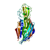
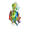
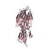
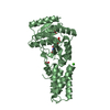
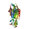
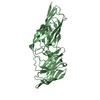
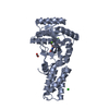
| Title | LEUKOCIDIN F (HLGB) FROM STAPHYLOCOCCUS AUREUS WITH PHOSPHOCHOLINE BOUND | ||||||
 Components Components | LEUKOCIDIN F SUBUNIT | ||||||
 Keywords Keywords | TOXIN / LEUKOTOXIN / HEMOLYSIN / PORE-FORMING TOXIN | ||||||
| Function / homology |  Function and homology information Function and homology information | ||||||
| Biological species |   Staphylococcus aureus (bacteria) Staphylococcus aureus (bacteria) | ||||||
| Method |  X-RAY DIFFRACTION / SYNCHROTRON / MOLECULAR REPLACEMENT / Resolution: 1.9 Å X-RAY DIFFRACTION / SYNCHROTRON / MOLECULAR REPLACEMENT / Resolution: 1.9 Å | ||||||
 Authors Authors | Olson, R. / Nariya, H. / Yokota, K. / Kamio, Y. / Gouaux, J.E. | ||||||
 Citation Citation | Journal: Nat.Struct.Biol. / Year: 1999 Title: Crystal structure of staphylococcal LukF delineates conformational changes accompanying formation of a transmembrane channel. Authors: Olson, R. / Nariya, H. / Yokota, K. / Kamio, Y. / Gouaux, E. | ||||||
| History |
|
- Structure visualization
| Structure viewer | Molecule: MolmilJmol/JSmol |
|---|
- Downloads & links
-Download
| PDBx/mmCIF format | 3lkf.cif.gz | 76.1 KB | Display | PDBx/mmCIF format |
|---|---|---|---|---|
| PDB format | pdb3lkf.ent.gz | 56.8 KB | Display | PDB format |
| PDBx/mmJSON format | 3lkf.json.gz | Tree view | PDBx/mmJSON format | |
| Others |  Other downloads Other downloads |
-Validation report
| Summary document | 3lkf_validation.pdf.gz | 375.7 KB | Display | wwPDB validaton report |
|---|---|---|---|---|
| Full document | 3lkf_full_validation.pdf.gz | 376.9 KB | Display | |
| Data in XML | 3lkf_validation.xml.gz | 7.4 KB | Display | |
| Data in CIF | 3lkf_validation.cif.gz | 12.3 KB | Display | |
| Arichive directory | https://data.pdbj.org/pub/pdb/validation_reports/lk/3lkfftp://data.pdbj.org/pub/pdb/validation_reports/lk/3lkf | HTTPS FTP |
-Related structure data


-Links
 PDBj
PDBj
- Assembly
| Deposited unit | 
| ||||||||
|---|---|---|---|---|---|---|---|---|---|
| 1 |
| ||||||||
| Unit cell |
|
-Components
| #1: Protein | Mass: 34090.719 Da / Num. of mol.: 1 / Fragment: WATER-SOLUBLE MONOMER Source method: isolated from a genetically manipulated source Source: (gene. exp.) Staphylococcus aureus (bacteria) / Strain: SMITH 5R / Gene: LUKF / Plasmid: PTRCLUKF / Gene (production host): LUKF / Production host: |
|---|---|
| #2: Chemical | ChemComp-PC /   Mass: 184.151 Da / Num. of mol.: 1 / Source method: obtained synthetically / Formula: C5H15NO4P Mass: 184.151 Da / Num. of mol.: 1 / Source method: obtained synthetically / Formula: C5H15NO4P |
| #3: Water | ChemComp-HOH /  Mass: 18.015 Da / Num. of mol.: 253 / Source method: isolated from a natural source / Formula: H2O Mass: 18.015 Da / Num. of mol.: 253 / Source method: isolated from a natural source / Formula: H2O |
-Experimental details
-Experiment
| Experiment | Method: X-RAY DIFFRACTION / Number of used crystals: 1 |
|---|
- Sample preparation
| Crystal | Density Matthews: 2.11 Å3/Da / Density % sol: 45 % | ||||||||||||||||||||||||||||||||||||
|---|---|---|---|---|---|---|---|---|---|---|---|---|---|---|---|---|---|---|---|---|---|---|---|---|---|---|---|---|---|---|---|---|---|---|---|---|---|
| Crystal grow | pH: 8.5 / Details: pH 8.5 | ||||||||||||||||||||||||||||||||||||
| Crystal | *PLUS | ||||||||||||||||||||||||||||||||||||
| Crystal grow | *PLUS Temperature: 20 ℃ / pH: 7.5 / Method: vapor diffusion | ||||||||||||||||||||||||||||||||||||
| Components of the solutions | *PLUS
|
-Data collection
| Diffraction | Mean temperature: 110 K |
|---|---|
| Diffraction source | Source: SYNCHROTRON / Site: APS  / Beamline: 14-BM-D / Wavelength: 0.9184 / Beamline: 14-BM-D / Wavelength: 0.9184 |
| Detector | Type: ADSC / Detector: CCD / Date: Jun 1, 1998 / Details: MIRRORS |
| Radiation | Monochromatic (M) / Laue (L): M / Scattering type: x-ray |
| Radiation wavelength | Wavelength: 0.9184 Å / Relative weight: 1 |
| Reflection | Resolution: 1.8→20 Å / Num. obs: 26850 / % possible obs: 97.2 % / Observed criterion σ(I): 0 / Redundancy: 3.5 % / Rsym value: 0.049 |
| Reflection shell | Resolution: 1.8→1.86 Å / Rsym value: 0.176 / % possible all: 86 |
| Reflection | *PLUS Rmerge(I) obs: 0.049 |
| Reflection shell | *PLUS % possible obs: 86 % / Rmerge(I) obs: 0.176 |
- Processing
| Software |
| ||||||||||||||||||||||||||||||||||||||||||||||||||||||||||||
|---|---|---|---|---|---|---|---|---|---|---|---|---|---|---|---|---|---|---|---|---|---|---|---|---|---|---|---|---|---|---|---|---|---|---|---|---|---|---|---|---|---|---|---|---|---|---|---|---|---|---|---|---|---|---|---|---|---|---|---|---|---|
| Refinement | Method to determine structure: MOLECULAR REPLACEMENT Starting model: PREVIOUSLY DETERMINED STRUCTURE Resolution: 1.9→6 Å / σ(F): 2
| ||||||||||||||||||||||||||||||||||||||||||||||||||||||||||||
| Displacement parameters | Biso mean: 11.01 Å2 | ||||||||||||||||||||||||||||||||||||||||||||||||||||||||||||
| Refinement step | Cycle: LAST / Resolution: 1.9→6 Å
| ||||||||||||||||||||||||||||||||||||||||||||||||||||||||||||
| Refine LS restraints |
| ||||||||||||||||||||||||||||||||||||||||||||||||||||||||||||
| Xplor file |
| ||||||||||||||||||||||||||||||||||||||||||||||||||||||||||||
| Software | *PLUS Name: X-PLOR / Version: 3.851 / Classification: refinement | ||||||||||||||||||||||||||||||||||||||||||||||||||||||||||||
| Refinement | *PLUS Num. reflection obs: 26815 | ||||||||||||||||||||||||||||||||||||||||||||||||||||||||||||
| Solvent computation | *PLUS | ||||||||||||||||||||||||||||||||||||||||||||||||||||||||||||
| Displacement parameters | *PLUS Biso mean: 11.017 Å2 | ||||||||||||||||||||||||||||||||||||||||||||||||||||||||||||
| Refine LS restraints | *PLUS
|
