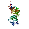
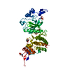
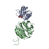
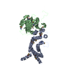
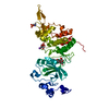
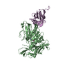
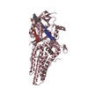
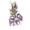
dual-specificity kinase / negative regulation of calcineurin-NFAT signaling cascade / smoothened signaling pathway / intrinsic apoptotic signaling pathway in response to DNA damage by p53 class mediator / positive regulation of glycogen biosynthetic process / ubiquitin ligase complex / protein serine/threonine/tyrosine kinase activity / regulation of signal transduction by p53 class mediator / manganese ion binding / protein tyrosine kinase activity ...dual-specificity kinase / negative regulation of calcineurin-NFAT signaling cascade / smoothened signaling pathway / intrinsic apoptotic signaling pathway in response to DNA damage by p53 class mediator / positive regulation of glycogen biosynthetic process / ubiquitin ligase complex / protein serine/threonine/tyrosine kinase activity / regulation of signal transduction by p53 class mediator / manganese ion binding / protein tyrosine kinase activity / Regulation of TP53 Activity through Phosphorylation / cytoskeleton / protein phosphorylation / ribonucleoprotein complex / protein serine kinase activity / protein serine/threonine kinase activity / DNA damage response / magnesium ion binding / nucleoplasm / ATP binding / nucleus / cytosol / cytoplasm Similarity search - Function
DYRK / Dual specificity tyrosine-phosphorylation-regulated kinase / Trypsin Inhibitor V; Chain A / : / Phosphorylase Kinase; domain 1 / Phosphorylase Kinase; domain 1 / Transferase(Phosphotransferase) domain 1 / Transferase(Phosphotransferase); domain 1 / Serine/threonine-protein kinase, active site / Serine/Threonine protein kinases active-site signature. ...DYRK / Dual specificity tyrosine-phosphorylation-regulated kinase / Trypsin Inhibitor V; Chain A / : / Phosphorylase Kinase; domain 1 / Phosphorylase Kinase; domain 1 / Transferase(Phosphotransferase) domain 1 / Transferase(Phosphotransferase); domain 1 / Serine/threonine-protein kinase, active site / Serine/Threonine protein kinases active-site signature. / Protein kinase domain / Serine/Threonine protein kinases, catalytic domain / Protein kinase, ATP binding site / Protein kinases ATP-binding region signature. / Protein kinase domain profile. / Protein kinase domain / Protein kinase-like domain superfamily / 2-Layer Sandwich / Orthogonal Bundle / Mainly Alpha / Alpha Beta Similarity search - Domain/homology
In the structure databanks used in Yorodumi, some data are registered as the other names, "COVID-19 virus" and "2019-nCoV". Here are the details of the virus and the list of structure data.
Jan 31, 2019. EMDB accession codes are about to change! (news from PDBe EMDB page)
EMDB accession codes are about to change! (news from PDBe EMDB page)
The allocation of 4 digits for EMDB accession codes will soon come to an end. Whilst these codes will remain in use, new EMDB accession codes will include an additional digit and will expand incrementally as the available range of codes is exhausted. The current 4-digit format prefixed with “EMD-” (i.e. EMD-XXXX) will advance to a 5-digit format (i.e. EMD-XXXXX), and so on. It is currently estimated that the 4-digit codes will be depleted around Spring 2019, at which point the 5-digit format will come into force.
The EM Navigator/Yorodumi systems omit the EMD- prefix.
Related info.:Q: What is EMD? / ID/Accession-code notation in Yorodumi/EM Navigator
Yorodumi is a browser for structure data from EMDB, PDB, SASBDB, etc.
This page is also the successor to EM Navigator detail page, and also detail information page/front-end page for Omokage search.
The word "yorodu" (or yorozu) is an old Japanese word meaning "ten thousand". "mi" (miru) is to see.
Related info.:EMDB / PDB / SASBDB / Comparison of 3 databanks / Yorodumi Search / Aug 31, 2016. New EM Navigator & Yorodumi / Yorodumi Papers / Jmol/JSmol / Function and homology information / Changes in new EM Navigator and Yorodumi
 Movie
Movie Controller

 Yorodumi
 Open data
 Basic information
 Components
 Keywords
 Function and homology information
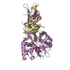
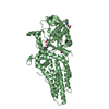
 Homo sapiens (human)
 X-RAY DIFFRACTION /
  Authors
 Citation
 Structure visualization
 Downloads & links
 Other downloads
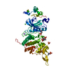
 PDBj
PDBj Assembly
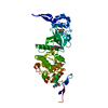

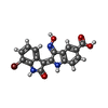
 Mass: 400.183 Da / Num. of mol.: 1 / Source method: obtained synthetically / Formula: C17H10BrN3O4
 Mass: 35.453 Da / Num. of mol.: 4 / Source method: obtained synthetically / Formula: Cl
 Mass: 18.015 Da / Num. of mol.: 107 / Source method: isolated from a natural source / Formula: H2O
 Sample preparation
 / Beamline: I03 / Wavelength: 0.9802 Å
 Processing