 Movie
Movie Controller

[English] 日本語
 Yorodumi
- PDB-3il5: Structure of E. faecalis FabH in complex with 2-({4-bromo-3-[(die... -
+ Open data

- Basic information
| Entry | Database: PDB / ID: 3il5 | ||||||
|---|---|---|---|---|---|---|---|
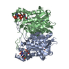
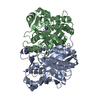
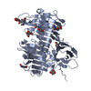
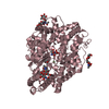
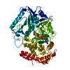
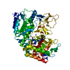
| Title | Structure of E. faecalis FabH in complex with 2-({4-bromo-3-[(diethylamino)sulfonyl]benzoyl}amino)benzoic acid | ||||||
 Components Components | 3-oxoacyl-[acyl-carrier-protein] synthase 3 | ||||||
 Keywords Keywords | TRANSFERASE / FabH / fatty acid biosynthesis / antibiotic / Acyltransferase / Cytoplasm / Lipid synthesis / Multifunctional enzyme | ||||||
| Function / homology |  Function and homology information Function and homology informationbeta-ketoacyl-[acyl-carrier-protein] synthase III / beta-ketoacyl-acyl-carrier-protein synthase III activity / 3-oxoacyl-[acyl-carrier-protein] synthase activity / fatty acid biosynthetic process / cytoplasm Similarity search - Function | ||||||
| Biological species |   Enterococcus faecalis (bacteria) Enterococcus faecalis (bacteria) | ||||||
| Method |  X-RAY DIFFRACTION / MOLECULAR REPLACEMENT / Resolution: 2.6 Å X-RAY DIFFRACTION / MOLECULAR REPLACEMENT / Resolution: 2.6 Å | ||||||
 Authors Authors | Gajiwala, K.S. / Margosiak, S. / Lu, J. / Cortez, J. / Su, Y. / Nie, Z. / Appelt, K. | ||||||
 Citation Citation | Journal: Febs Lett. / Year: 2009 Title: Crystal structures of bacterial FabH suggest a molecular basis for the substrate specificity of the enzyme. Authors: Gajiwala, K.S. / Margosiak, S. / Lu, J. / Cortez, J. / Su, Y. / Nie, Z. / Appelt, K. | ||||||
| History |
|
- Structure visualization
| Structure viewer | Molecule: MolmilJmol/JSmol |
|---|
- Downloads & links
-Download
| PDBx/mmCIF format | 3il5.cif.gz | 254.8 KB | Display | PDBx/mmCIF format |
|---|---|---|---|---|
| PDB format | pdb3il5.ent.gz | 206.9 KB | Display | PDB format |
| PDBx/mmJSON format | 3il5.json.gz | Tree view | PDBx/mmJSON format | |
| Others |  Other downloads Other downloads |
-Validation report
| Arichive directory | https://data.pdbj.org/pub/pdb/validation_reports/il/3il5ftp://data.pdbj.org/pub/pdb/validation_reports/il/3il5 | HTTPS FTP |
|---|
-Related structure data
| Related structure data | 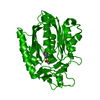 3il3C  3il4C  3il6C 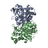 3il7C 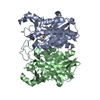 3il9SC C: citing same article ( S: Starting model for refinement |
|---|---|
| Similar structure data |
-Links
 PDBj
PDBj

- Assembly
| Deposited unit | 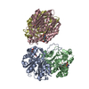
| ||||||||
|---|---|---|---|---|---|---|---|---|---|
| 1 | 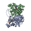
| ||||||||
| 2 | 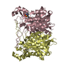
| ||||||||
| Unit cell |
|
-Components
| #1: Protein | Mass: 37663.496 Da / Num. of mol.: 4 Source method: isolated from a genetically manipulated source Source: (gene. exp.) Enterococcus faecalis (bacteria) / Gene: EF_2885, fabH / Production host: References: UniProt: Q820T1, beta-ketoacyl-[acyl-carrier-protein] synthase III #2: Chemical | ChemComp-B82 /   Mass: 455.323 Da / Num. of mol.: 4 / Source method: obtained synthetically / Formula: C18H19BrN2O5S Mass: 455.323 Da / Num. of mol.: 4 / Source method: obtained synthetically / Formula: C18H19BrN2O5S#3: Water | ChemComp-HOH / |  Mass: 18.015 Da / Num. of mol.: 128 / Source method: isolated from a natural source / Formula: H2O Mass: 18.015 Da / Num. of mol.: 128 / Source method: isolated from a natural source / Formula: H2O |
|---|
-Experimental details
-Experiment
| Experiment | Method: X-RAY DIFFRACTION / Number of used crystals: 1 |
|---|
- Sample preparation
| Crystal | Density Matthews: 2.12 Å3/Da / Density % sol: 42.1 % |
|---|---|
| Crystal grow | Temperature: 288 K / Method: vapor diffusion, hanging drop / pH: 6.5 Details: 0.2 M lithium sulfate, 0.1 M Bis-Tris, pH 6.5, 25% PEG3350, VAPOR DIFFUSION, HANGING DROP, temperature 288K |
-Data collection
| Diffraction | Mean temperature: 120 K |
|---|---|
| Diffraction source | Source: ROTATING ANODE / Type: ENRAF-NONIUS FR591 / Wavelength: 1.5418 Å |
| Detector | Type: MAR scanner 345 mm plate / Detector: IMAGE PLATE / Date: Jan 1, 2004 / Details: mirrors |
| Radiation | Monochromator: mirrors / Protocol: SINGLE WAVELENGTH / Monochromatic (M) / Laue (L): M / Scattering type: x-ray |
| Radiation wavelength | Wavelength: 1.5418 Å / Relative weight: 1 |
| Reflection | Resolution: 2.6→25 Å / Num. all: 38916 / Num. obs: 38358 / % possible obs: 98.6 % / Observed criterion σ(F): 1 / Observed criterion σ(I): 1 / Redundancy: 3.8 % / Rmerge(I) obs: 0.12 / Rsym value: 0.12 / Net I/σ(I): 11.3 |
| Reflection shell | Resolution: 2.6→2.69 Å / Redundancy: 3.6 % / Rmerge(I) obs: 0.338 / Mean I/σ(I) obs: 3.1 / Num. unique all: 3767 / Rsym value: 0.338 / % possible all: 97.3 |
- Processing
| Software |
| ||||||||||||||||||||||||||||||||||||
|---|---|---|---|---|---|---|---|---|---|---|---|---|---|---|---|---|---|---|---|---|---|---|---|---|---|---|---|---|---|---|---|---|---|---|---|---|---|
| Refinement | Method to determine structure: MOLECULAR REPLACEMENT Starting model: 3IL9 Resolution: 2.6→25 Å / Occupancy max: 1 / Occupancy min: 1 / σ(F): 0 / σ(I): 0 / Stereochemistry target values: Engh & Huber
| ||||||||||||||||||||||||||||||||||||
| Displacement parameters | Biso max: 123.34 Å2 / Biso mean: 43.762 Å2 / Biso min: 12.37 Å2
| ||||||||||||||||||||||||||||||||||||
| Refine analyze | Luzzati coordinate error obs: 0.3 Å / Luzzati d res low obs: 5 Å / Luzzati sigma a obs: 0.39 Å | ||||||||||||||||||||||||||||||||||||
| Refinement step | Cycle: LAST / Resolution: 2.6→25 Å
| ||||||||||||||||||||||||||||||||||||
| Refine LS restraints |
| ||||||||||||||||||||||||||||||||||||
| LS refinement shell | Resolution: 2.6→2.76 Å / Rfactor Rfree error: 0.016
|
