 Movie
Movie Controller

[English] 日本語
 Yorodumi
- PDB-1os1: Structure of Phosphoenolpyruvate Carboxykinase complexed with ATP... -
+ Open data

- Basic information
| Entry | Database: PDB / ID: 1os1 | |||||||||
|---|---|---|---|---|---|---|---|---|---|---|
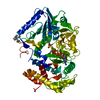
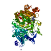
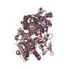
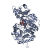
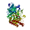
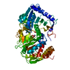
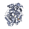
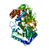
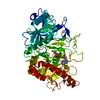
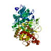
| Title | Structure of Phosphoenolpyruvate Carboxykinase complexed with ATP,Mg, Ca and pyruvate. | |||||||||
 Components Components | Phosphoenolpyruvate carboxykinase [ATP] | |||||||||
 Keywords Keywords | LYASE / enzyme mechanism / phosphotransfer / calcium / activation | |||||||||
| Function / homology |  Function and homology information Function and homology informationphosphoenolpyruvate carboxykinase (ATP) / phosphoenolpyruvate carboxykinase (ATP) activity / gluconeogenesis / calcium ion binding / magnesium ion binding / ATP binding / cytosol Similarity search - Function | |||||||||
| Biological species |  | |||||||||
| Method |  X-RAY DIFFRACTION / SYNCHROTRON / MOLECULAR REPLACEMENT / Resolution: 1.8 Å X-RAY DIFFRACTION / SYNCHROTRON / MOLECULAR REPLACEMENT / Resolution: 1.8 Å | |||||||||
 Authors Authors | Sudom, A. / Walters, R. / Pastushok, L. / Goldie, D. / Prasad, L. / Delbaere, L.T. / Goldie, H. | |||||||||
 Citation Citation | Journal: J.BACTERIOL. / Year: 2003 Title: Mechanisms of activation of phosphoenolpyruvate carboxykinase from Escherichia coli by Ca2+ and of desensitization by trypsin. Authors: Sudom, A. / Walters, R. / Pastushok, L. / Goldie, D. / Prasad, L. / Delbaere, L.T. / Goldie, H. | |||||||||
| History |
|
- Structure visualization
| Structure viewer | Molecule: MolmilJmol/JSmol |
|---|
- Downloads & links
-Download
| PDBx/mmCIF format | 1os1.cif.gz | 124 KB | Display | PDBx/mmCIF format |
|---|---|---|---|---|
| PDB format | pdb1os1.ent.gz | 93.2 KB | Display | PDB format |
| PDBx/mmJSON format | 1os1.json.gz | Tree view | PDBx/mmJSON format | |
| Others |  Other downloads Other downloads |
-Validation report
| Arichive directory | https://data.pdbj.org/pub/pdb/validation_reports/os/1os1ftp://data.pdbj.org/pub/pdb/validation_reports/os/1os1 | HTTPS FTP |
|---|
-Related structure data
| Related structure data |  1aqlS S: Starting model for refinement |
|---|---|
| Similar structure data |
-Links
 PDBj
PDBj

- Assembly
| Deposited unit | 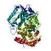
| ||||||||
|---|---|---|---|---|---|---|---|---|---|
| 1 |
| ||||||||
| Unit cell |
|
-Components
-Protein , 1 types, 1 molecules A
| #1: Protein | Mass: 59709.176 Da / Num. of mol.: 1 / Source method: isolated from a natural source / Source: (natural) References: UniProt: P22259, phosphoenolpyruvate carboxykinase (ATP) |
|---|
-Non-polymers , 5 types, 264 molecules 


| #2: Chemical | ChemComp-MG /  Mass: 24.305 Da / Num. of mol.: 1 / Source method: obtained synthetically / Formula: Mg Mass: 24.305 Da / Num. of mol.: 1 / Source method: obtained synthetically / Formula: Mg |
|---|---|
| #3: Chemical | ChemComp-CA /  Mass: 40.078 Da / Num. of mol.: 1 / Source method: obtained synthetically / Formula: Ca Mass: 40.078 Da / Num. of mol.: 1 / Source method: obtained synthetically / Formula: Ca |
| #4: Chemical | ChemComp-ATP /  Mass: 507.181 Da / Num. of mol.: 1 / Source method: obtained synthetically / Formula: C10H16N5O13P3 / Comment: ATP, energy-carrying molecule*YM Mass: 507.181 Da / Num. of mol.: 1 / Source method: obtained synthetically / Formula: C10H16N5O13P3 / Comment: ATP, energy-carrying molecule*YM |
| #5: Chemical | ChemComp-PYR /  Mass: 88.062 Da / Num. of mol.: 1 / Source method: obtained synthetically / Formula: C3H4O3 Mass: 88.062 Da / Num. of mol.: 1 / Source method: obtained synthetically / Formula: C3H4O3 |
| #6: Water | ChemComp-HOH / Mass: 18.015 Da / Num. of mol.: 260 / Source method: isolated from a natural source / Formula: H2O |
-Experimental details
-Experiment
| Experiment | Method: X-RAY DIFFRACTION / Number of used crystals: 1 |
|---|
- Sample preparation
| Crystal | Density Matthews: 2.26 Å3/Da / Density % sol: 45.12 % | ||||||||||||||||||||||||||||||||||||||||||||||||||||||||||||||||||||||||||||||||||||||||||||||||||
|---|---|---|---|---|---|---|---|---|---|---|---|---|---|---|---|---|---|---|---|---|---|---|---|---|---|---|---|---|---|---|---|---|---|---|---|---|---|---|---|---|---|---|---|---|---|---|---|---|---|---|---|---|---|---|---|---|---|---|---|---|---|---|---|---|---|---|---|---|---|---|---|---|---|---|---|---|---|---|---|---|---|---|---|---|---|---|---|---|---|---|---|---|---|---|---|---|---|---|---|
| Crystal grow | Temperature: 294 K / Method: vapor diffusion, hanging drop / pH: 4.8 Details: ADP, Calcium Chloride, Magnesium Chloride, PEP, EDTA, ammonium acetate, sodium acetate buffer, dithiothreitol, PEG 4000, pH 4.8, VAPOR DIFFUSION, HANGING DROP, temperature 294K | ||||||||||||||||||||||||||||||||||||||||||||||||||||||||||||||||||||||||||||||||||||||||||||||||||
| Crystal grow | *PLUS Temperature: 21 ℃ / Method: vapor diffusion, hanging drop | ||||||||||||||||||||||||||||||||||||||||||||||||||||||||||||||||||||||||||||||||||||||||||||||||||
| Components of the solutions | *PLUS
|
-Data collection
| Diffraction source | Source: SYNCHROTRON / Site: Photon Factory  / Beamline: BL-18B / Wavelength: 1 Å / Beamline: BL-18B / Wavelength: 1 Å |
|---|---|
| Detector | Type: FUJI / Detector: IMAGE PLATE / Date: Dec 14, 1998 |
| Radiation | Protocol: SINGLE WAVELENGTH / Monochromatic (M) / Laue (L): M / Scattering type: x-ray |
| Radiation wavelength | Wavelength: 1 Å / Relative weight: 1 |
| Reflection | Resolution: 1.8→40 Å / Num. all: 41488 / Num. obs: 41488 / % possible obs: 78.3 % / Observed criterion σ(I): 0 / Redundancy: 2.84 % / Rsym value: 0.037 |
| Reflection | *PLUS Lowest resolution: 10 Å / Rmerge(I) obs: 0.037 |
| Reflection shell | *PLUS Highest resolution: 1.8 Å / Lowest resolution: 1.86 Å / % possible obs: 69.4 % / Rmerge(I) obs: 0.45 / Mean I/σ(I) obs: 1.13 |
- Processing
| Software |
| |||||||||||||||||||||
|---|---|---|---|---|---|---|---|---|---|---|---|---|---|---|---|---|---|---|---|---|---|---|
| Refinement | Method to determine structure: MOLECULAR REPLACEMENT Starting model: PDB ENTRY 1AQL Resolution: 1.8→10 Å / Isotropic thermal model: Isotropic / σ(F): 0 / Stereochemistry target values: Engh & Huber / Details: Simulated annealing
| |||||||||||||||||||||
| Displacement parameters | Biso mean: 29.6 Å2 | |||||||||||||||||||||
| Refinement step | Cycle: LAST / Resolution: 1.8→10 Å
| |||||||||||||||||||||
| Refine LS restraints |
| |||||||||||||||||||||
| Refinement | *PLUS Highest resolution: 1.8 Å / Lowest resolution: 10 Å / % reflection Rfree: 5 % / Rfactor Rfree: 0.22 / Rfactor Rwork: 0.18 | |||||||||||||||||||||
| Solvent computation | *PLUS | |||||||||||||||||||||
| Displacement parameters | *PLUS | |||||||||||||||||||||
| Refine LS restraints | *PLUS
| |||||||||||||||||||||
| LS refinement shell | *PLUS Highest resolution: 1.8 Å / Lowest resolution: 1.86 Å / Rfactor Rfree: 0.3 / Rfactor Rwork: 0.3 / Num. reflection obs: 2917 |
