 Movie
Movie Controller

[English] 日本語
 Yorodumi
- PDB-3feg: Crystal structure of human choline kinase beta in complex with ph... -
+ Open data

- Basic information
| Entry | Database: PDB / ID: 3feg | ||||||
|---|---|---|---|---|---|---|---|
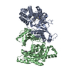
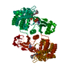
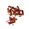
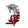
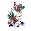
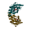
| Title | Crystal structure of human choline kinase beta in complex with phosphorylated hemicholinium-3 and adenosine nucleotide | ||||||
 Components Components | Choline/ethanolamine kinase | ||||||
 Keywords Keywords | TRANSFERASE / non-protein kinase / choline kinase / Structural Genomics Consortium / SGC / hemicholinium-3 / phosphorylation / Kinase / Phosphoprotein | ||||||
| Function / homology |  Function and homology information Function and homology informationethanolamine kinase / choline kinase / Synthesis of PE / ethanolamine kinase activity / choline kinase activity / CDP-choline pathway / phosphatidylethanolamine biosynthetic process / Synthesis of PC / ATP binding / cytoplasm / cytosol Similarity search - Function | ||||||
| Biological species |  Homo sapiens (human) Homo sapiens (human) | ||||||
| Method |  X-RAY DIFFRACTION / SYNCHROTRON / MOLECULAR REPLACEMENT / molecular replacement / Resolution: 1.302 Å X-RAY DIFFRACTION / SYNCHROTRON / MOLECULAR REPLACEMENT / molecular replacement / Resolution: 1.302 Å | ||||||
 Authors Authors | Hong, B.S. / Tempel, W. / Rabeh, W.M. / MacKenzie, F. / Arrowsmith, C.H. / Edwards, A.M. / Bountra, C. / Weigelt, J. / Bochkarev, A. / Park, H.W. / Structural Genomics Consortium (SGC) | ||||||
 Citation Citation | Journal: J.Biol.Chem. / Year: 2010 Title: Crystal structures of human choline kinase isoforms in complex with hemicholinium-3: single amino acid near the active site influences inhibitor sensitivity. Authors: Hong, B.S. / Allali-Hassani, A. / Tempel, W. / Finerty, P.J. / Mackenzie, F. / Dimov, S. / Vedadi, M. / Park, H.W. | ||||||
| History |
|
- Structure visualization
| Structure viewer | Molecule: MolmilJmol/JSmol |
|---|
- Downloads & links
-Download
| PDBx/mmCIF format | 3feg.cif.gz | 178.9 KB | Display | PDBx/mmCIF format |
|---|---|---|---|---|
| PDB format | pdb3feg.ent.gz | 138.1 KB | Display | PDB format |
| PDBx/mmJSON format | 3feg.json.gz | Tree view | PDBx/mmJSON format | |
| Others |  Other downloads Other downloads |
-Validation report
| Arichive directory | https://data.pdbj.org/pub/pdb/validation_reports/fe/3fegftp://data.pdbj.org/pub/pdb/validation_reports/fe/3feg | HTTPS FTP |
|---|
-Related structure data
| Related structure data |  3g15C  3lq3C  2ig7S S: Starting model for refinement C: citing same article ( |
|---|---|
| Similar structure data |


-Links
 PDBj
PDBj


- Assembly
| Deposited unit | 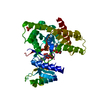
| ||||||||
|---|---|---|---|---|---|---|---|---|---|
| 1 | 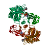
| ||||||||
| Unit cell |
|
-Components
-Protein , 1 types, 1 molecules A
| #1: Protein | Mass: 44002.613 Da / Num. of mol.: 1 Source method: isolated from a genetically manipulated source Source: (gene. exp.) Homo sapiens (human) / Gene: CHKB, CHETK, CHKL / Plasmid: pET28a-LIC / Production host:  References: UniProt: Q9Y259, choline kinase, ethanolamine kinase |
|---|
-Non-polymers , 7 types, 305 molecules 


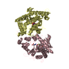
| #2: Chemical | ChemComp-HC7 / ( Mass: 494.518 Da / Num. of mol.: 1 / Source method: obtained synthetically / Formula: C24H35N2O7P Mass: 494.518 Da / Num. of mol.: 1 / Source method: obtained synthetically / Formula: C24H35N2O7P | ||
|---|---|---|---|
| #3: Chemical | ChemComp-SO4 /  Mass: 96.063 Da / Num. of mol.: 1 / Source method: obtained synthetically / Formula: SO4 Mass: 96.063 Da / Num. of mol.: 1 / Source method: obtained synthetically / Formula: SO4 | ||
| #4: Chemical | ChemComp-ADP /  Mass: 427.201 Da / Num. of mol.: 1 / Source method: obtained synthetically / Formula: C10H15N5O10P2 / Comment: ADP, energy-carrying molecule*YM Mass: 427.201 Da / Num. of mol.: 1 / Source method: obtained synthetically / Formula: C10H15N5O10P2 / Comment: ADP, energy-carrying molecule*YM | ||
| #5: Chemical | ChemComp-AMP /  Mass: 347.221 Da / Num. of mol.: 1 / Source method: obtained synthetically / Formula: C10H14N5O7P / Comment: AMP*YM Mass: 347.221 Da / Num. of mol.: 1 / Source method: obtained synthetically / Formula: C10H14N5O7P / Comment: AMP*YM | ||
| #6: Chemical | ChemComp-MG /  Mass: 24.305 Da / Num. of mol.: 1 / Source method: obtained synthetically / Formula: Mg Mass: 24.305 Da / Num. of mol.: 1 / Source method: obtained synthetically / Formula: Mg | ||
| #7: Chemical |  Num. of mol.: 2 / Source method: obtained synthetically Num. of mol.: 2 / Source method: obtained synthetically#8: Water | ChemComp-HOH / | Mass: 18.015 Da / Num. of mol.: 298 / Source method: isolated from a natural source / Formula: H2O |
-Details
| Nonpolymer details | FOR THE LIGAND HC7 THE STEREO-CENTER AT THE "2" POSITION IS SPECIFIED AS (2S), HOWEVER CAN BE (2R) ...FOR THE LIGAND HC7 THE STEREO-CENTER AT THE "2" POSITION IS SPECIFIED AS (2S), HOWEVER CAN BE (2R) OR (2S) AS THE MORPHOLINI |
|---|
-Experimental details
-Experiment
| Experiment | Method: X-RAY DIFFRACTION / Number of used crystals: 1 |
|---|
- Sample preparation
| Crystal | Density Matthews: 2.24 Å3/Da / Density % sol: 45.18 % |
|---|---|
| Crystal grow | Temperature: 291 K / Method: vapor diffusion / pH: 6.5 Details: protein buffer: 0.01M Tris pH 8, 0.5M sodium chloride, 0.005M magnesium chloride, 0.01M DTT, 0.003M hemicholinium-3, 0.005M ADP; precipitant: 0.1M sodium cacodylate, 30% PEG-4000, 0.2M ...Details: protein buffer: 0.01M Tris pH 8, 0.5M sodium chloride, 0.005M magnesium chloride, 0.01M DTT, 0.003M hemicholinium-3, 0.005M ADP; precipitant: 0.1M sodium cacodylate, 30% PEG-4000, 0.2M ammonium sulfate, VAPOR DIFFUSION, temperature 291K |
-Data collection
| Diffraction | Mean temperature: 100 K | ||||||||||||||||||||||||||||||||||||||||||||||||||||||||||||||||||
|---|---|---|---|---|---|---|---|---|---|---|---|---|---|---|---|---|---|---|---|---|---|---|---|---|---|---|---|---|---|---|---|---|---|---|---|---|---|---|---|---|---|---|---|---|---|---|---|---|---|---|---|---|---|---|---|---|---|---|---|---|---|---|---|---|---|---|---|
| Diffraction source | Source: SYNCHROTRON / Site: APS  / Beamline: 23-ID-B / Wavelength: 0.97243 Å / Beamline: 23-ID-B / Wavelength: 0.97243 Å | ||||||||||||||||||||||||||||||||||||||||||||||||||||||||||||||||||
| Detector | Type: MARMOSAIC 300 mm CCD / Detector: CCD / Date: Mar 6, 2008 | ||||||||||||||||||||||||||||||||||||||||||||||||||||||||||||||||||
| Radiation | Protocol: SINGLE WAVELENGTH / Monochromatic (M) / Laue (L): M / Scattering type: x-ray | ||||||||||||||||||||||||||||||||||||||||||||||||||||||||||||||||||
| Radiation wavelength | Wavelength: 0.97243 Å / Relative weight: 1 | ||||||||||||||||||||||||||||||||||||||||||||||||||||||||||||||||||
| Reflection | Resolution: 1.3→30 Å / Num. obs: 92472 / % possible obs: 97.6 % / Redundancy: 3.6 % / Rmerge(I) obs: 0.04 / Χ2: 1.578 / Net I/σ(I): 14.4 | ||||||||||||||||||||||||||||||||||||||||||||||||||||||||||||||||||
| Reflection shell |
|
-Phasing
| Phasing | Method: molecular replacement |
|---|
- Processing
| Software |
| |||||||||||||||||||||||||||||||||||||||||||||||||||||||||||||||||||||||||||||||||||||||||||||||||||||||||||||||||||||||||||||||||||||||||||||||||||
|---|---|---|---|---|---|---|---|---|---|---|---|---|---|---|---|---|---|---|---|---|---|---|---|---|---|---|---|---|---|---|---|---|---|---|---|---|---|---|---|---|---|---|---|---|---|---|---|---|---|---|---|---|---|---|---|---|---|---|---|---|---|---|---|---|---|---|---|---|---|---|---|---|---|---|---|---|---|---|---|---|---|---|---|---|---|---|---|---|---|---|---|---|---|---|---|---|---|---|---|---|---|---|---|---|---|---|---|---|---|---|---|---|---|---|---|---|---|---|---|---|---|---|---|---|---|---|---|---|---|---|---|---|---|---|---|---|---|---|---|---|---|---|---|---|---|---|---|---|
| Refinement | Method to determine structure: MOLECULAR REPLACEMENT Starting model: pdb entry 2ig7 Resolution: 1.302→28.296 Å / Cor.coef. Fo:Fc: 0.97 / Cor.coef. Fo:Fc free: 0.959 / WRfactor Rfree: 0.201 / WRfactor Rwork: 0.162 / SU B: 1.51 / SU ML: 0.029 / Cross valid method: THROUGHOUT / σ(F): 0 / ESU R: 0.05 / ESU R Free: 0.05 / Stereochemistry target values: Engh & Huber Details: HYDROGENS HAVE BEEN ADDED IN THE RIDING POSITIONS. Hemicholinium restraints were calculated by the PRODRG server.
| |||||||||||||||||||||||||||||||||||||||||||||||||||||||||||||||||||||||||||||||||||||||||||||||||||||||||||||||||||||||||||||||||||||||||||||||||||
| Solvent computation | Ion probe radii: 0.8 Å / Shrinkage radii: 0.8 Å / VDW probe radii: 1.2 Å / Solvent model: MASK BULK SOLVENT | |||||||||||||||||||||||||||||||||||||||||||||||||||||||||||||||||||||||||||||||||||||||||||||||||||||||||||||||||||||||||||||||||||||||||||||||||||
| Displacement parameters | Biso mean: 14.43 Å2
| |||||||||||||||||||||||||||||||||||||||||||||||||||||||||||||||||||||||||||||||||||||||||||||||||||||||||||||||||||||||||||||||||||||||||||||||||||
| Refinement step | Cycle: LAST / Resolution: 1.302→28.296 Å
| |||||||||||||||||||||||||||||||||||||||||||||||||||||||||||||||||||||||||||||||||||||||||||||||||||||||||||||||||||||||||||||||||||||||||||||||||||
| Refine LS restraints |
| |||||||||||||||||||||||||||||||||||||||||||||||||||||||||||||||||||||||||||||||||||||||||||||||||||||||||||||||||||||||||||||||||||||||||||||||||||
| LS refinement shell | Refine-ID: X-RAY DIFFRACTION / Total num. of bins used: 20
|
