 Movie
Movie Controller

[English] 日本語
 Yorodumi
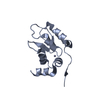
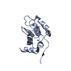
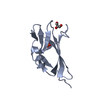
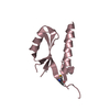
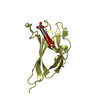
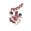
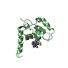
- PDB-3f7g: Structure of the BIR domain from ML-IAP bound to a peptidomimetic -
+ Open data

- Basic information
| Entry | Database: PDB / ID: 3f7g | ||||||
|---|---|---|---|---|---|---|---|
| Title | Structure of the BIR domain from ML-IAP bound to a peptidomimetic | ||||||
 Components Components | Baculoviral IAP repeat-containing protein 7 | ||||||
 Keywords Keywords | APOPTOSIS / ZINC BINDING / PEPTIDE COMPLEX / APOPTOSIS INHIBITION / PEPTIDOMIMETIC / SMALL MOLECULE / DRUG DESIGN / Metal-binding / Nucleus / Zinc-finger | ||||||
| Function / homology |  Function and homology information Function and homology informationregulation of natural killer cell apoptotic process / Regulation of MITF-M-dependent genes involved in apoptosis / cysteine-type endopeptidase inhibitor activity involved in apoptotic process / lens development in camera-type eye / cysteine-type endopeptidase inhibitor activity / negative regulation of tumor necrosis factor-mediated signaling pathway / positive regulation of protein ubiquitination / RING-type E3 ubiquitin transferase / positive regulation of JNK cascade / ubiquitin-protein transferase activity ...regulation of natural killer cell apoptotic process / Regulation of MITF-M-dependent genes involved in apoptosis / cysteine-type endopeptidase inhibitor activity involved in apoptotic process / lens development in camera-type eye / cysteine-type endopeptidase inhibitor activity / negative regulation of tumor necrosis factor-mediated signaling pathway / positive regulation of protein ubiquitination / RING-type E3 ubiquitin transferase / positive regulation of JNK cascade / ubiquitin-protein transferase activity / ubiquitin protein ligase activity / regulation of cell population proliferation / regulation of apoptotic process / regulation of cell cycle / protein ubiquitination / apoptotic process / centrosome / negative regulation of apoptotic process / enzyme binding / Golgi apparatus / zinc ion binding / nucleoplasm / nucleus / cytosol / cytoplasm Similarity search - Function | ||||||
| Biological species |  Homo sapiens (human) Homo sapiens (human) | ||||||
| Method |  X-RAY DIFFRACTION / SYNCHROTRON / difference Fourier / Resolution: 2.3 Å X-RAY DIFFRACTION / SYNCHROTRON / difference Fourier / Resolution: 2.3 Å | ||||||
 Authors Authors | Franklin, M.C. / Fairbrother, W.J. / Cohen, F. | ||||||
 Citation Citation | Journal: J.Med.Chem. / Year: 2009 Title: Orally bioavailable antagonists of inhibitor of apoptosis proteins based on an azabicyclooctane scaffold Authors: Cohen, F. / Alicke, B. / Elliott, L.O. / Flygare, J.A. / Goncharov, T. / Keteltas, S.F. / Franklin, M.C. / Frankovitz, S. / Stephan, J.P. / Tsui, V. / Vucic, D. / Wong, H. / Fairbrother, W.J. #1: Journal: ACS Chem. Biol. / Year: 2006Title: Design, synthesis, and biological activity of a potent Smac mimetic that sensitizes cancer cells to apoptosis by antagonizing IAPs. Authors: Zobel, K. / Wang, L. / Varfolomeev, E. / Franklin, M.C. / Elliott, L.O. / Wallweber, H.J. / Okawa, D.C. / Flygare, J.A. / Vucic, D. / Fairbrother, W.J. / Deshayes, K. #2: Journal: Biochem. J. / Year: 2005Title: Engineering ML-IAP to produce an extraordinarily potent caspase-9 inhibitor: implications for Smac-dependent anti-apoptotic activity of ML-IAP. Authors: Vucic, D. / Franklin, M.C. / Wallweber, H.J. / Das, K. / Eckelman, B.P. / Shin, H. / Elliott, L.O. / Deshayes, K. / Salvesen, G.S. / Fairbrother, W.J. #3: Journal: Biochemistry / Year: 2003Title: Structure and function analysis of peptide antagonists of melanoma inhibitor of apoptosis (ML-IAP) Authors: Franklin, M.C. / Kadkhodayan, S. / Ackerly, H. / Alexandru, D. / Distefano, M.D. / Elliott, L.O. / Flygare, J.A. / Vucic, D. / Deshayes, K. / Fairbrother, W.J. | ||||||
| History |
|
- Structure visualization
| Structure viewer | Molecule: MolmilJmol/JSmol |
|---|
- Downloads & links
-Download
| PDBx/mmCIF format | 3f7g.cif.gz | 123.5 KB | Display | PDBx/mmCIF format |
|---|---|---|---|---|
| PDB format | pdb3f7g.ent.gz | 94.3 KB | Display | PDB format |
| PDBx/mmJSON format | 3f7g.json.gz | Tree view | PDBx/mmJSON format | |
| Others |  Other downloads Other downloads |
-Validation report
| Arichive directory | https://data.pdbj.org/pub/pdb/validation_reports/f7/3f7gftp://data.pdbj.org/pub/pdb/validation_reports/f7/3f7g | HTTPS FTP |
|---|
-Related structure data
| Related structure data | 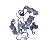 3f7hC 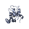 3f7iC  1oxnS S: Starting model for refinement C: citing same article ( |
|---|---|
| Similar structure data |


-Links
 PDBj
PDBj


- Assembly
| Deposited unit | 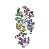
| ||||||||
|---|---|---|---|---|---|---|---|---|---|
| 1 | 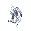
| ||||||||
| 2 | 
| ||||||||
| 3 | 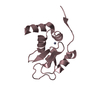
| ||||||||
| 4 | 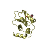
| ||||||||
| 5 | 
| ||||||||
| 6 | 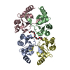
| ||||||||
| Unit cell |
| ||||||||
| Details | Authors state that each asymmetric unit contains five biological units, each containing one molecule of ML-IAP with either a peptide or a peptidomimetic bound at the active site. Chains A-D form a (not biologically relevant) tetramer in which molecule A binds a peptide consisting of part of the N-terminal unstructured region of molecule D; molecule B similarly binds part of molecule C; molecule C binds molecule B; and molecule D binds molecule A. The biologically relevant complex is formed by ML-IAP molecule E binding the peptidomimetic (ligand 389). |
-Components
| #1: Protein | Mass: 15765.368 Da / Num. of mol.: 5 / Fragment: BIR domain, residues 63-179 Source method: isolated from a genetically manipulated source Source: (gene. exp.) Homo sapiens (human)Gene: also known as KIAP OR MLIAP OR LIVIN, BIRC7, KIAP, LIVIN, MLIAP, RNF50, UNQ5800/PRO19607/PRO21344 Plasmid: pET15B / Production host:  #2: Chemical | ChemComp-ZN /   Mass: 65.409 Da / Num. of mol.: 5 / Source method: obtained synthetically / Formula: Zn Mass: 65.409 Da / Num. of mol.: 5 / Source method: obtained synthetically / Formula: Zn#3: Chemical | ChemComp-P33 / |   Mass: 326.383 Da / Num. of mol.: 1 / Source method: obtained synthetically / Formula: C14H30O8 / Comment: precipitant*YM Mass: 326.383 Da / Num. of mol.: 1 / Source method: obtained synthetically / Formula: C14H30O8 / Comment: precipitant*YM#4: Chemical | ChemComp-389 / | 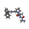  Mass: 464.600 Da / Num. of mol.: 1 / Source method: obtained synthetically / Formula: C27H36N4O3 Mass: 464.600 Da / Num. of mol.: 1 / Source method: obtained synthetically / Formula: C27H36N4O3#5: Water | ChemComp-HOH / |  Mass: 18.015 Da / Num. of mol.: 270 / Source method: isolated from a natural source / Formula: H2O Mass: 18.015 Da / Num. of mol.: 270 / Source method: isolated from a natural source / Formula: H2O |
|---|
-Experimental details
-Experiment
| Experiment | Method: X-RAY DIFFRACTION / Number of used crystals: 1 |
|---|
- Sample preparation
| Crystal | Density Matthews: 2.44 Å3/Da / Density % sol: 49.64 % |
|---|---|
| Crystal grow | Temperature: 298 K / Method: vapor diffusion, sitting drop / pH: 5 Details: SODIUM ACETATE, PEG 300, DTT, pH 5.0, VAPOR DIFFUSION, SITTING DROP, temperature 298K |
-Data collection
| Diffraction | Mean temperature: 100 K |
|---|---|
| Diffraction source | Source: SYNCHROTRON / Site: SSRL  / Beamline: BL7-1 / Wavelength: 1.08 Å / Beamline: BL7-1 / Wavelength: 1.08 Å |
| Detector | Type: MAR scanner 345 mm plate / Detector: IMAGE PLATE / Date: Nov 19, 2002 |
| Radiation | Monochromator: Vertical focusing mirror; single crystal (Si111) bent monochromator (horizontal focusing). Protocol: SINGLE WAVELENGTH / Monochromatic (M) / Laue (L): M / Scattering type: x-ray |
| Radiation wavelength | Wavelength: 1.08 Å / Relative weight: 1 |
| Reflection | Resolution: 2.3→30 Å / Num. all: 33128 / Num. obs: 32773 / % possible obs: 98.9 % / Observed criterion σ(F): 0 / Observed criterion σ(I): -3 / Redundancy: 1.9 % / Biso Wilson estimate: 40.6 Å2 / Rmerge(I) obs: 0.061 / Rsym value: 0.061 / Net I/σ(I): 11.8 |
| Reflection shell | Resolution: 2.3→2.38 Å / Redundancy: 1.9 % / Rmerge(I) obs: 0.431 / Mean I/σ(I) obs: 1.9 / Num. unique all: 3304 / Rsym value: 0.431 / % possible all: 98.3 |
- Processing
| Software |
| ||||||||||||||||||||||||||||||||||||||||||||||||||||||||||||||||||||||||||||||||||||||||||||||||||||||||||||||||||||||||||||||||||||||||||||||||||||||
|---|---|---|---|---|---|---|---|---|---|---|---|---|---|---|---|---|---|---|---|---|---|---|---|---|---|---|---|---|---|---|---|---|---|---|---|---|---|---|---|---|---|---|---|---|---|---|---|---|---|---|---|---|---|---|---|---|---|---|---|---|---|---|---|---|---|---|---|---|---|---|---|---|---|---|---|---|---|---|---|---|---|---|---|---|---|---|---|---|---|---|---|---|---|---|---|---|---|---|---|---|---|---|---|---|---|---|---|---|---|---|---|---|---|---|---|---|---|---|---|---|---|---|---|---|---|---|---|---|---|---|---|---|---|---|---|---|---|---|---|---|---|---|---|---|---|---|---|---|---|---|---|
| Refinement | Method to determine structure: difference Fourier Starting model: PDB entry 1OXN Resolution: 2.3→20 Å / Cor.coef. Fo:Fc: 0.957 / Cor.coef. Fo:Fc free: 0.936 / SU B: 9.463 / SU ML: 0.128 / TLS residual ADP flag: LIKELY RESIDUAL / Cross valid method: THROUGHOUT / σ(F): 0 / ESU R: 0.217 / ESU R Free: 0.182 / Stereochemistry target values: MAXIMUM LIKELIHOOD / Details: HYDROGENS HAVE BEEN ADDED IN THE RIDING POSITIONS
| ||||||||||||||||||||||||||||||||||||||||||||||||||||||||||||||||||||||||||||||||||||||||||||||||||||||||||||||||||||||||||||||||||||||||||||||||||||||
| Solvent computation | Ion probe radii: 0.8 Å / Shrinkage radii: 0.8 Å / VDW probe radii: 1.4 Å / Solvent model: BABINET MODEL WITH MASK | ||||||||||||||||||||||||||||||||||||||||||||||||||||||||||||||||||||||||||||||||||||||||||||||||||||||||||||||||||||||||||||||||||||||||||||||||||||||
| Displacement parameters | Biso mean: 33.954 Å2
| ||||||||||||||||||||||||||||||||||||||||||||||||||||||||||||||||||||||||||||||||||||||||||||||||||||||||||||||||||||||||||||||||||||||||||||||||||||||
| Refinement step | Cycle: LAST / Resolution: 2.3→20 Å
| ||||||||||||||||||||||||||||||||||||||||||||||||||||||||||||||||||||||||||||||||||||||||||||||||||||||||||||||||||||||||||||||||||||||||||||||||||||||
| Refine LS restraints |
| ||||||||||||||||||||||||||||||||||||||||||||||||||||||||||||||||||||||||||||||||||||||||||||||||||||||||||||||||||||||||||||||||||||||||||||||||||||||
| LS refinement shell | Resolution: 2.3→2.359 Å / Total num. of bins used: 20
| ||||||||||||||||||||||||||||||||||||||||||||||||||||||||||||||||||||||||||||||||||||||||||||||||||||||||||||||||||||||||||||||||||||||||||||||||||||||
| Refinement TLS params. | Method: refined / Refine-ID: X-RAY DIFFRACTION
| ||||||||||||||||||||||||||||||||||||||||||||||||||||||||||||||||||||||||||||||||||||||||||||||||||||||||||||||||||||||||||||||||||||||||||||||||||||||
| Refinement TLS group |
|
