 Movie
Movie Controller

[English] 日本語
 Yorodumi
- PDB-3ejb: Crystal Structure of P450BioI in complex with tetradecanoic acid ... -
+ Open data

- Basic information
| Entry | Database: PDB / ID: 3ejb | ||||||
|---|---|---|---|---|---|---|---|
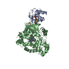
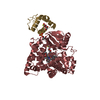
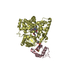
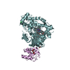
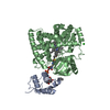
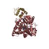
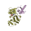
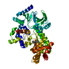
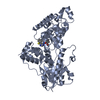
| Title | Crystal Structure of P450BioI in complex with tetradecanoic acid ligated Acyl Carrier Protein | ||||||
 Components Components |
| ||||||
 Keywords Keywords | Oxidoreductase/Lipid Transport / Protein-Protein Complex / Cytochrome P450 Fold / Carrier Protein / 4-Helix Bundle / Fatty acid biosynthesis / Lipid synthesis / Phosphopantetheine / Biotin biosynthesis / Heme / Iron / Metal-binding / Monooxygenase / Oxidoreductase / Oxidoreductase-Lipid Transport COMPLEX | ||||||
| Function / homology |  Function and homology information Function and homology informationpimeloyl-[acyl-carrier protein] synthase / biotin biosynthetic process / lipid A biosynthetic process / lipid biosynthetic process / phosphopantetheine binding / acyl binding / oxidoreductase activity, acting on paired donors, with incorporation or reduction of molecular oxygen / acyl carrier activity / monooxygenase activity / fatty acid biosynthetic process ...pimeloyl-[acyl-carrier protein] synthase / biotin biosynthetic process / lipid A biosynthetic process / lipid biosynthetic process / phosphopantetheine binding / acyl binding / oxidoreductase activity, acting on paired donors, with incorporation or reduction of molecular oxygen / acyl carrier activity / monooxygenase activity / fatty acid biosynthetic process / iron ion binding / response to xenobiotic stimulus / heme binding / lipid binding / membrane / cytoplasm / cytosol Similarity search - Function | ||||||
| Biological species |  | ||||||
| Method |  X-RAY DIFFRACTION / SYNCHROTRON / FOURIER SYNTHESIS / Resolution: 2 Å X-RAY DIFFRACTION / SYNCHROTRON / FOURIER SYNTHESIS / Resolution: 2 Å | ||||||
 Authors Authors | Cryle, M.J. / Schlichting, I. | ||||||
 Citation Citation | Journal: Proc.Natl.Acad.Sci.Usa / Year: 2008 Title: Structural insights from a P450 Carrier Protein complex reveal how specificity is achieved in the P450(BioI) ACP complex. Authors: Cryle, M.J. / Schlichting, I. | ||||||
| History |
|
- Structure visualization
| Structure viewer | Molecule: MolmilJmol/JSmol |
|---|
- Downloads & links
-Download
| PDBx/mmCIF format | 3ejb.cif.gz | 405.9 KB | Display | PDBx/mmCIF format |
|---|---|---|---|---|
| PDB format | pdb3ejb.ent.gz | 329.9 KB | Display | PDB format |
| PDBx/mmJSON format | 3ejb.json.gz | Tree view | PDBx/mmJSON format | |
| Others |  Other downloads Other downloads |
-Validation report
| Arichive directory | https://data.pdbj.org/pub/pdb/validation_reports/ej/3ejbftp://data.pdbj.org/pub/pdb/validation_reports/ej/3ejb | HTTPS FTP |
|---|
-Related structure data


-Links
 PDBj
PDBj

- Assembly
| Deposited unit | 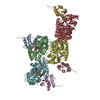
| ||||||||
|---|---|---|---|---|---|---|---|---|---|
| 1 | 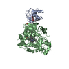
| ||||||||
| 2 | 
| ||||||||
| 3 | 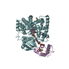
| ||||||||
| 4 | 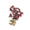
| ||||||||
| Unit cell |
| ||||||||
| Details | heterodimers are formed by chain A and B, C and D, E and F, G and H. |
-Components
-Protein , 2 types, 8 molecules ACEGBDFH
| #1: Protein | Mass: 10685.630 Da / Num. of mol.: 4 Source method: isolated from a genetically manipulated source Source: (gene. exp.) #2: Protein | Mass: 45988.352 Da / Num. of mol.: 4 Source method: isolated from a genetically manipulated source Source: (gene. exp.) References: UniProt: P53554, Oxidoreductases; Acting on paired donors, with incorporation or reduction of molecular oxygen |
|---|
-Sugars , 1 types, 8 molecules 
| #4: Sugar | ChemComp-HTG /  Type: D-saccharide / Mass: 294.408 Da / Num. of mol.: 8 / Source method: obtained synthetically / Formula: C13H26O5S / Comment: detergent*YM Type: D-saccharide / Mass: 294.408 Da / Num. of mol.: 8 / Source method: obtained synthetically / Formula: C13H26O5S / Comment: detergent*YM |
|---|
-Non-polymers , 4 types, 1348 molecules 


| #3: Chemical | ChemComp-ZMP /  Mass: 568.704 Da / Num. of mol.: 4 / Source method: obtained synthetically / Formula: C25H49N2O8PS Mass: 568.704 Da / Num. of mol.: 4 / Source method: obtained synthetically / Formula: C25H49N2O8PS#5: Chemical | ChemComp-HEM /  Mass: 616.487 Da / Num. of mol.: 4 / Source method: obtained synthetically / Formula: C34H32FeN4O4 Mass: 616.487 Da / Num. of mol.: 4 / Source method: obtained synthetically / Formula: C34H32FeN4O4#6: Chemical |  Mass: 35.453 Da / Num. of mol.: 2 / Source method: obtained synthetically / Formula: Cl Mass: 35.453 Da / Num. of mol.: 2 / Source method: obtained synthetically / Formula: Cl#7: Water | ChemComp-HOH / | Mass: 18.015 Da / Num. of mol.: 1338 / Source method: isolated from a natural source / Formula: H2O |
|---|
-Details
| Has protein modification | Y |
|---|
-Experimental details
-Experiment
| Experiment | Method: X-RAY DIFFRACTION / Number of used crystals: 1 |
|---|
- Sample preparation
| Crystal | Density Matthews: 2.54 Å3/Da / Density % sol: 51.49 % |
|---|---|
| Crystal grow | Temperature: 277 K / Method: vapor diffusion, hanging drop / pH: 6.5 Details: 0.1 M Na HEPES, 0.25 M NaCl, 0.15 M Li2SO4, 19% PEG 4000, 0.2% n-heptyl b-D-thioglucopyranoside, pH 6.5, VAPOR DIFFUSION, HANGING DROP, temperature 277K |
-Data collection
| Diffraction | Mean temperature: 100 K |
|---|---|
| Diffraction source | Source: SYNCHROTRON / Site: SLS  / Beamline: X10SA / Wavelength: 0.98089 Å / Beamline: X10SA / Wavelength: 0.98089 Å |
| Detector | Type: MARMOSAIC 225 mm CCD / Detector: CCD / Date: Mar 8, 2008 |
| Radiation | Monochromator: SI(111) / Protocol: SINGLE WAVELENGTH / Monochromatic (M) / Laue (L): M / Scattering type: x-ray |
| Radiation wavelength | Wavelength: 0.98089 Å / Relative weight: 1 |
| Reflection | Resolution: 2→50 Å / Num. all: 149113 / Num. obs: 144447 / % possible obs: 96.9 % / Observed criterion σ(F): 0 / Observed criterion σ(I): 0 / Redundancy: 7.9 % / Biso Wilson estimate: 25.9 Å2 / Rsym value: 0.0604 / Net I/σ(I): 21.8 |
| Reflection shell | Resolution: 2→2.05 Å / Redundancy: 7.4 % / Mean I/σ(I) obs: 7.4 / Rsym value: 0.33 / % possible all: 94.9 |
- Processing
| Software |
| ||||||||||||||||||||||||||||||||||||||||||||||||||||||||||||||||||||||||||||||||||||||||||||||||||||||||||||||||||||||||||||||||||||||||||||||||||||||||||||||||||||||||||
|---|---|---|---|---|---|---|---|---|---|---|---|---|---|---|---|---|---|---|---|---|---|---|---|---|---|---|---|---|---|---|---|---|---|---|---|---|---|---|---|---|---|---|---|---|---|---|---|---|---|---|---|---|---|---|---|---|---|---|---|---|---|---|---|---|---|---|---|---|---|---|---|---|---|---|---|---|---|---|---|---|---|---|---|---|---|---|---|---|---|---|---|---|---|---|---|---|---|---|---|---|---|---|---|---|---|---|---|---|---|---|---|---|---|---|---|---|---|---|---|---|---|---|---|---|---|---|---|---|---|---|---|---|---|---|---|---|---|---|---|---|---|---|---|---|---|---|---|---|---|---|---|---|---|---|---|---|---|---|---|---|---|---|---|---|---|---|---|---|---|---|---|
| Refinement | Method to determine structure: FOURIER SYNTHESIS Starting model: SAD Structure Resolution: 2→20 Å / Cor.coef. Fo:Fc: 0.936 / Cor.coef. Fo:Fc free: 0.904 / SU B: 4.965 / SU ML: 0.141 / Cross valid method: THROUGHOUT / ESU R: 0.215 / ESU R Free: 0.187 / Stereochemistry target values: MAXIMUM LIKELIHOOD / Details: HYDROGENS HAVE BEEN ADDED IN THE RIDING POSITIONS
| ||||||||||||||||||||||||||||||||||||||||||||||||||||||||||||||||||||||||||||||||||||||||||||||||||||||||||||||||||||||||||||||||||||||||||||||||||||||||||||||||||||||||||
| Solvent computation | Ion probe radii: 0.8 Å / Shrinkage radii: 0.8 Å / VDW probe radii: 1.2 Å / Solvent model: BABINET MODEL WITH MASK | ||||||||||||||||||||||||||||||||||||||||||||||||||||||||||||||||||||||||||||||||||||||||||||||||||||||||||||||||||||||||||||||||||||||||||||||||||||||||||||||||||||||||||
| Displacement parameters | Biso mean: 28.83 Å2
| ||||||||||||||||||||||||||||||||||||||||||||||||||||||||||||||||||||||||||||||||||||||||||||||||||||||||||||||||||||||||||||||||||||||||||||||||||||||||||||||||||||||||||
| Refinement step | Cycle: LAST / Resolution: 2→20 Å
| ||||||||||||||||||||||||||||||||||||||||||||||||||||||||||||||||||||||||||||||||||||||||||||||||||||||||||||||||||||||||||||||||||||||||||||||||||||||||||||||||||||||||||
| Refine LS restraints |
| ||||||||||||||||||||||||||||||||||||||||||||||||||||||||||||||||||||||||||||||||||||||||||||||||||||||||||||||||||||||||||||||||||||||||||||||||||||||||||||||||||||||||||
| LS refinement shell | Resolution: 2→2.05 Å / Total num. of bins used: 20
|
