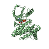
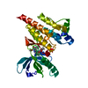
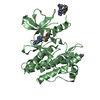
Entry Database : PDB / ID : 3bz3Title Crystal Structure Analysis of Focal Adhesion Kinase with a Methanesulfonamide Diaminopyrimidine Inhibitor Focal adhesion kinase 1 Keywords / / / / / / / / / Function / homology Function Domain/homology Component
/ / / / / / / / / / / / / / / / / / / / / / / / / / / / / / / / / / / / / / / / / / / / / / / / / / / / / / / / / / / / / / / / / / / / / / / / / / / / / / / / / / / / / / / / / / / / / / / / / / / / / / / / / / / / / / / / / / / / / / / / / / / / / / / / / / / / / / / / / Biological species Homo sapiens (human)Method / / / Resolution : 2.2 Å Authors Vajdos, F. / Marr, E. Journal : Cancer Res. / Year : 2008Title : Antitumor activity and pharmacology of a selective focal adhesion kinase inhibitor, PF-562,271.Authors: Roberts, W.G. / Ung, E. / Whalen, P. / Cooper, B. / Hulford, C. / Autry, C. / Richter, D. / Emerson, E. / Lin, J. / Kath, J. / Coleman, K. / Yao, L. / Martinez-Alsina, L. / Lorenzen, M. / ... Authors : Roberts, W.G. / Ung, E. / Whalen, P. / Cooper, B. / Hulford, C. / Autry, C. / Richter, D. / Emerson, E. / Lin, J. / Kath, J. / Coleman, K. / Yao, L. / Martinez-Alsina, L. / Lorenzen, M. / Berliner, M. / Luzzio, M. / Patel, N. / Schmitt, E. / LaGreca, S. / Jani, J. / Wessel, M. / Marr, E. / Griffor, M. / Vajdos, F. History Deposition Jan 17, 2008 Deposition site / Processing site Revision 1.0 Apr 1, 2008 Provider / Type Revision 1.1 Jul 13, 2011 Group Revision 1.2 Oct 25, 2017 Group / Category Revision 1.3 Oct 30, 2024 Group Data collection / Database references ... Data collection / Database references / Derived calculations / Structure summary Category chem_comp_atom / chem_comp_bond ... chem_comp_atom / chem_comp_bond / database_2 / pdbx_entry_details / pdbx_modification_feature / struct_site Item _database_2.pdbx_DOI / _database_2.pdbx_database_accession ... _database_2.pdbx_DOI / _database_2.pdbx_database_accession / _struct_site.pdbx_auth_asym_id / _struct_site.pdbx_auth_comp_id / _struct_site.pdbx_auth_seq_id
Show all Show less
 Movie
Movie Controller

 Yorodumi
 Open data
 Basic information
 Components
 Keywords
 Function and homology information
 Homo sapiens (human)
 X-RAY DIFFRACTION /
  Authors
 Citation
 Structure visualization
 Downloads & links
 Other downloads
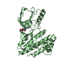


 PDBj
PDBj


 Assembly

 Spodoptera frugiperda (fall armyworm) / Strain (production host): Sf9
 Mass: 507.489 Da / Num. of mol.: 1 / Source method: obtained synthetically / Formula: C21H20F3N7O3S
 Mass: 18.015 Da / Num. of mol.: 281 / Source method: isolated from a natural source / Formula: H2O
 Sample preparation
 Processing