 Movie
Movie Controller

[English] 日本語
 Yorodumi
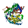
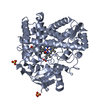
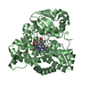
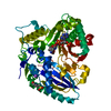
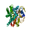
- PDB-3b30: Crystal Structure of F. graminearum TRI101 complexed with Ethyl C... -
+ Open data

- Basic information
| Entry | Database: PDB / ID: 3b30 | ||||||
|---|---|---|---|---|---|---|---|
| Title | Crystal Structure of F. graminearum TRI101 complexed with Ethyl Coenzyme A | ||||||
 Components Components | Trichothecene 3-O-acetyltransferase | ||||||
 Keywords Keywords | TRANSFERASE / acetyltransferase / BAHD superfamily / trichothecene / deoxynivalenol / T-2 / acetyl CoA / Fusarium / TRI101 | ||||||
| Function / homology |  Function and homology information Function and homology information | ||||||
| Biological species |  Gibberella zeae (fungus) Gibberella zeae (fungus) | ||||||
| Method |  X-RAY DIFFRACTION / SYNCHROTRON / MOLECULAR REPLACEMENT / Resolution: 1.97 Å X-RAY DIFFRACTION / SYNCHROTRON / MOLECULAR REPLACEMENT / Resolution: 1.97 Å | ||||||
 Authors Authors | Garvey, G.S. / Rayment, I. | ||||||
 Citation Citation | Journal: J.Biol.Chem. / Year: 2008 Title: Structural and Functional Characterization of the TRI101 Trichothecene 3-O-Acetyltransferase from Fusarium sporotrichioides and Fusarium graminearum: KINETIC INSIGHTS TO COMBATING FUSARIUM HEAD BLIGHT Authors: Garvey, G.S. / McCormick, S.P. / Rayment, I. | ||||||
| History |
|
- Structure visualization
| Structure viewer | Molecule: MolmilJmol/JSmol |
|---|
- Downloads & links
-Download
| PDBx/mmCIF format | 3b30.cif.gz | 107.4 KB | Display | PDBx/mmCIF format |
|---|---|---|---|---|
| PDB format | pdb3b30.ent.gz | 80.6 KB | Display | PDB format |
| PDBx/mmJSON format | 3b30.json.gz | Tree view | PDBx/mmJSON format | |
| Others |  Other downloads Other downloads |
-Validation report
| Arichive directory | https://data.pdbj.org/pub/pdb/validation_reports/b3/3b30ftp://data.pdbj.org/pub/pdb/validation_reports/b3/3b30 | HTTPS FTP |
|---|
-Related structure data
| Related structure data |  2rktC  2rkvC 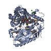 2zbaSC  3b2sC S: Starting model for refinement C: citing same article ( |
|---|---|
| Similar structure data |


-Links
 PDBj
PDBj

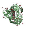
- Assembly
| Deposited unit | 
| ||||||||||||
|---|---|---|---|---|---|---|---|---|---|---|---|---|---|
| 1 |
| ||||||||||||
| Unit cell |
| ||||||||||||
| Components on special symmetry positions |
|
-Components
| #1: Protein | Mass: 49449.086 Da / Num. of mol.: 1 Source method: isolated from a genetically manipulated source Source: (gene. exp.) Gibberella zeae (fungus) / Strain: PH-1 / Gene: Tri101 / Plasmid details: rTEV cleavable N terminal His Tag / Plasmid: pET31b / Production host:  |
|---|---|
| #2: Chemical | ChemComp-ETB /   Mass: 721.443 Da / Num. of mol.: 1 / Source method: obtained synthetically / Formula: C20H34N7O16P3 Mass: 721.443 Da / Num. of mol.: 1 / Source method: obtained synthetically / Formula: C20H34N7O16P3 |
| #3: Chemical | ChemComp-MPO /   Mass: 209.263 Da / Num. of mol.: 1 / Source method: obtained synthetically / Formula: C7H15NO4S / Comment: pH buffer*YM Mass: 209.263 Da / Num. of mol.: 1 / Source method: obtained synthetically / Formula: C7H15NO4S / Comment: pH buffer*YM |
| #4: Water | ChemComp-HOH /  Mass: 18.015 Da / Num. of mol.: 327 / Source method: isolated from a natural source / Formula: H2O Mass: 18.015 Da / Num. of mol.: 327 / Source method: isolated from a natural source / Formula: H2O |
-Experimental details
-Experiment
| Experiment | Method: X-RAY DIFFRACTION / Number of used crystals: 1 |
|---|
- Sample preparation
| Crystal | Density Matthews: 3.12 Å3/Da / Density % sol: 60.58 % |
|---|---|
| Crystal grow | Temperature: 277 K / Method: vapor diffusion, hanging drop / pH: 6.9 Details: 2.1M sodium malonate, 100 mM 3-N-morpholino propanesulfonic acid, pH 6.9, VAPOR DIFFUSION, HANGING DROP, temperature 277K |
-Data collection
| Diffraction | Mean temperature: 100 K |
|---|---|
| Diffraction source | Source: SYNCHROTRON / Site: APS  / Beamline: 19-BM / Wavelength: 0.97907 Å / Beamline: 19-BM / Wavelength: 0.97907 Å |
| Detector | Detector: CCD |
| Radiation | Protocol: SINGLE WAVELENGTH / Monochromatic (M) / Laue (L): M / Scattering type: x-ray |
| Radiation wavelength | Wavelength: 0.97907 Å / Relative weight: 1 |
| Reflection | Resolution: 1.97→50 Å / Num. obs: 612294 / % possible obs: 99.9 % / Redundancy: 7.3 % / Biso Wilson estimate: 17.2 Å2 / Rmerge(I) obs: 0.079 / Net I/σ(I): 19.7 |
| Reflection shell | Resolution: 1.97→2.04 Å / Redundancy: 6.5 % / Rmerge(I) obs: 0.079 / Mean I/σ(I) obs: 19.7 / % possible all: 99.3 |
- Processing
| Software |
| ||||||||||||||||||||||||||||||||||||||||||||||||||||||||||||||||||||||||||||||||||||||||||||||||||||||||||||||||||||||||||||||||||||||||||||||||||||||||||||||||||||||||||
|---|---|---|---|---|---|---|---|---|---|---|---|---|---|---|---|---|---|---|---|---|---|---|---|---|---|---|---|---|---|---|---|---|---|---|---|---|---|---|---|---|---|---|---|---|---|---|---|---|---|---|---|---|---|---|---|---|---|---|---|---|---|---|---|---|---|---|---|---|---|---|---|---|---|---|---|---|---|---|---|---|---|---|---|---|---|---|---|---|---|---|---|---|---|---|---|---|---|---|---|---|---|---|---|---|---|---|---|---|---|---|---|---|---|---|---|---|---|---|---|---|---|---|---|---|---|---|---|---|---|---|---|---|---|---|---|---|---|---|---|---|---|---|---|---|---|---|---|---|---|---|---|---|---|---|---|---|---|---|---|---|---|---|---|---|---|---|---|---|---|---|---|
| Refinement | Method to determine structure: MOLECULAR REPLACEMENT Starting model: PDB entry 2ZBA Resolution: 1.97→35.14 Å / Cor.coef. Fo:Fc: 0.942 / Cor.coef. Fo:Fc free: 0.929 / SU B: 2.752 / SU ML: 0.08 / Cross valid method: THROUGHOUT / ESU R: 0.138 / ESU R Free: 0.125 / Stereochemistry target values: MAXIMUM LIKELIHOOD
| ||||||||||||||||||||||||||||||||||||||||||||||||||||||||||||||||||||||||||||||||||||||||||||||||||||||||||||||||||||||||||||||||||||||||||||||||||||||||||||||||||||||||||
| Solvent computation | Ion probe radii: 0.8 Å / Shrinkage radii: 0.8 Å / VDW probe radii: 1.2 Å / Solvent model: MASK | ||||||||||||||||||||||||||||||||||||||||||||||||||||||||||||||||||||||||||||||||||||||||||||||||||||||||||||||||||||||||||||||||||||||||||||||||||||||||||||||||||||||||||
| Displacement parameters | Biso mean: 14.887 Å2
| ||||||||||||||||||||||||||||||||||||||||||||||||||||||||||||||||||||||||||||||||||||||||||||||||||||||||||||||||||||||||||||||||||||||||||||||||||||||||||||||||||||||||||
| Refinement step | Cycle: LAST / Resolution: 1.97→35.14 Å
| ||||||||||||||||||||||||||||||||||||||||||||||||||||||||||||||||||||||||||||||||||||||||||||||||||||||||||||||||||||||||||||||||||||||||||||||||||||||||||||||||||||||||||
| Refine LS restraints |
| ||||||||||||||||||||||||||||||||||||||||||||||||||||||||||||||||||||||||||||||||||||||||||||||||||||||||||||||||||||||||||||||||||||||||||||||||||||||||||||||||||||||||||
| LS refinement shell | Resolution: 1.97→2.023 Å / Total num. of bins used: 20
|
