 Movie
Movie Controller


+ Open data

- Basic information
| Entry | Database: EMDB / ID: EMD-3872 | ||||||||||||
|---|---|---|---|---|---|---|---|---|---|---|---|---|---|
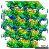
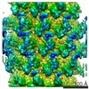
| Title | Cryo-electron tomogram of Ebola virus virus-like particles | ||||||||||||
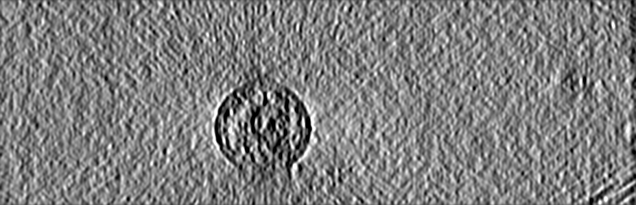
 Map data Map data | A 4x binned representative tomogram of recombinant virus-like particles from Ebola virus nucleoprotein, VP24, VP35, VP40. | ||||||||||||
 Sample Sample |
| ||||||||||||
| Biological species |   Ebola virus - Mayinga, Zaire, 1976 Ebola virus - Mayinga, Zaire, 1976 | ||||||||||||
| Method | electron tomography / cryo EM | ||||||||||||
 Authors Authors | Wan W / Kolesnikova L / Clarke M / Koehler A / Noda T / Becker S / Briggs JAG | ||||||||||||
| Funding support |  Germany, 3 items Germany, 3 items
| ||||||||||||
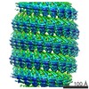
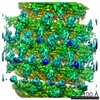
 Citation Citation | Journal: Nature / Year: 2017 Title: Structure and assembly of the Ebola virus nucleocapsid. Authors: William Wan / Larissa Kolesnikova / Mairi Clarke / Alexander Koehler / Takeshi Noda / Stephan Becker / John A G Briggs /   Abstract: Ebola and Marburg viruses are filoviruses: filamentous, enveloped viruses that cause haemorrhagic fever. Filoviruses are within the order Mononegavirales, which also includes rabies virus, measles ...Ebola and Marburg viruses are filoviruses: filamentous, enveloped viruses that cause haemorrhagic fever. Filoviruses are within the order Mononegavirales, which also includes rabies virus, measles virus, and respiratory syncytial virus. Mononegaviruses have non-segmented, single-stranded negative-sense RNA genomes that are encapsidated by nucleoprotein and other viral proteins to form a helical nucleocapsid. The nucleocapsid acts as a scaffold for virus assembly and as a template for genome transcription and replication. Insights into nucleoprotein-nucleoprotein interactions have been derived from structural studies of oligomerized, RNA-encapsidating nucleoprotein, and cryo-electron microscopy of nucleocapsid or nucleocapsid-like structures. There have been no high-resolution reconstructions of complete mononegavirus nucleocapsids. Here we apply cryo-electron tomography and subtomogram averaging to determine the structure of Ebola virus nucleocapsid within intact viruses and recombinant nucleocapsid-like assemblies. These structures reveal the identity and arrangement of the nucleocapsid components, and suggest that the formation of an extended α-helix from the disordered carboxy-terminal region of nucleoprotein-core links nucleoprotein oligomerization, nucleocapsid condensation, RNA encapsidation, and accessory protein recruitment. | ||||||||||||
| History |
|
- Structure visualization
| Movie |
 Movie viewer Movie viewer |
|---|---|
| Structure viewer | EM map: SurfViewMolmilJmol/JSmol |
| Supplemental images |

- Downloads & links
-EMDB archive
| Map data | emd_3872.map.gz | 246.5 MB | EMDB map data format | |
|---|---|---|---|---|
| Header (meta data) | emd-3872-v30.xmlemd-3872.xml | 18.4 KB 18.4 KB | Display Display | EMDB header |
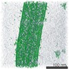
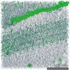
| Images | 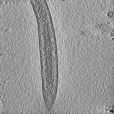 emd_3872.png emd_3872.png | 259 KB | ||
| Archive directory |  http://ftp.pdbj.org/pub/emdb/structures/EMD-3872ftp://ftp.pdbj.org/pub/emdb/structures/EMD-3872 http://ftp.pdbj.org/pub/emdb/structures/EMD-3872ftp://ftp.pdbj.org/pub/emdb/structures/EMD-3872 | HTTPS FTP |
-Validation report
| Summary document | emd_3872_validation.pdf.gz | 174.1 KB | Display | EMDB validaton report |
|---|---|---|---|---|
| Full document | emd_3872_full_validation.pdf.gz | 173.3 KB | Display | |
| Data in XML | emd_3872_validation.xml.gz | 3.7 KB | Display | |
| Arichive directory | https://ftp.pdbj.org/pub/emdb/validation_reports/EMD-3872ftp://ftp.pdbj.org/pub/emdb/validation_reports/EMD-3872 | HTTPS FTP |
-Related structure data
-Links
| EMDB pages | EMDB (EBI/PDBe) / EMDataResource |
|---|
-Map
| File | Download / File: emd_3872.map.gz / Format: CCP4 / Size: 491.7 MB / Type: IMAGE STORED AS SIGNED INTEGER (2 BYTES) | ||||||||||||||||||||||||||||||||||||||||||||||||||||||||||||||||||||
|---|---|---|---|---|---|---|---|---|---|---|---|---|---|---|---|---|---|---|---|---|---|---|---|---|---|---|---|---|---|---|---|---|---|---|---|---|---|---|---|---|---|---|---|---|---|---|---|---|---|---|---|---|---|---|---|---|---|---|---|---|---|---|---|---|---|---|---|---|---|
| Annotation | A 4x binned representative tomogram of recombinant virus-like particles from Ebola virus nucleoprotein, VP24, VP35, VP40. | ||||||||||||||||||||||||||||||||||||||||||||||||||||||||||||||||||||
| Projections & slices | Image control
Images are generated by Spider. generated in cubic-lattice coordinate | ||||||||||||||||||||||||||||||||||||||||||||||||||||||||||||||||||||
| Voxel size | X=Y=Z: 7.12 Å | ||||||||||||||||||||||||||||||||||||||||||||||||||||||||||||||||||||
| Density |
| ||||||||||||||||||||||||||||||||||||||||||||||||||||||||||||||||||||
| Symmetry | Space group: 1 | ||||||||||||||||||||||||||||||||||||||||||||||||||||||||||||||||||||
| Details | EMDB XML:
CCP4 map header:
| ||||||||||||||||||||||||||||||||||||||||||||||||||||||||||||||||||||
 Z (Sec.)
 Y (Row.)
 X (Col.)


-Supplemental data
- Sample components
-Entire : Ebola virus - Mayinga, Zaire, 1976
| Entire | Name: Ebola virus - Mayinga, Zaire, 1976 |
|---|---|
| Components |
|
-Supramolecule #1: Ebola virus - Mayinga, Zaire, 1976
| Supramolecule | Name: Ebola virus - Mayinga, Zaire, 1976 / type: virus / ID: 1 / Parent: 0 / Macromolecule list: all Details: Recombinantly expressed virus-like particles produced by expression of nucleoprotein, VP24, VP35, VP40. NCBI-ID: 128952 / Sci species name: Ebola virus - Mayinga, Zaire, 1976 / Virus type: VIRUS-LIKE PARTICLE / Virus isolate: STRAIN / Virus enveloped: Yes / Virus empty: No |
|---|---|
| Host system | Organism:  Homo sapiens (human) / Recombinant cell: HEK 293T Homo sapiens (human) / Recombinant cell: HEK 293T |
| Virus shell | Shell ID: 1 / Name: Nucleocapsid / Diameter: 280.0 Å |
-Macromolecule #1: Ebola virus nucleoprotein
| Macromolecule | Name: Ebola virus nucleoprotein / type: protein_or_peptide / ID: 1 / Enantiomer: LEVO |
|---|---|
| Source (natural) | Organism: Ebola virus - Mayinga, Zaire, 1976 |
| Recombinant expression | Organism: Homo sapiens (human) |
| Sequence | String: MDSRPQKIWM APSLTESDMD YHKILTAGLS VQQGIVRQRV IPVYQVNNLE EICQLIIQAF EAGVDFQES ADSFLLMLCL HHAYQGDYKL FLESGAVKYL EGHGFRFEVK KRDGVKRLEE L LPAVSSGK NIKRTLAAMP EEETTEANAG QFLSFASLFL PKLVVGEKAC ...String: MDSRPQKIWM APSLTESDMD YHKILTAGLS VQQGIVRQRV IPVYQVNNLE EICQLIIQAF EAGVDFQES ADSFLLMLCL HHAYQGDYKL FLESGAVKYL EGHGFRFEVK KRDGVKRLEE L LPAVSSGK NIKRTLAAMP EEETTEANAG QFLSFASLFL PKLVVGEKAC LEKVQRQIQV HA EQGLIQY PTAWQSVGHM MVIFRLMRTN FLIKFLLIHQ GMHMVAGHDA NDAVISNSVA QAR FSGLLI VKTVLDHILQ KTERGVRLHP LARTAKVKNE VNSFKAALSS LAKHGEYAPF ARLL NLSGV NNLEHGLFPQ LSAIALGVAT AHGSTLAGVN VGEQYQQLRE AATEAEKQLQ QYAES RELD HLGLDDQEKK ILMNFHQKKN EISFQQTNAM VTLRKERLAK LTEAITAASL PKTSGH YDD DDDIPFPGPI NDDDNPGHQD DDPTDSQDTT IPDVVVDPDD GSYGEYQSYS ENGMNAP DD LVLFDLDEDD EDTKPVPNRS TKGGQQKNSQ KGQHIEGRQT QSRPIQNVPG PHRTIHHA S APLTDNDRRN EPSGSTSPRM LTPINEEADP LDDADDETSS LPPLESDDEE QDRDGTSNR TPTVAPPAPV YRDHSEKKEL PQDEQQDQDH TQEARNQDSD NTQSEHSFEE MYRHILRSQG PFDAVLYYH MMKDEPVVFS TSDGKEYTYP DSLEEEYPPW LTEKEAMNEE NRFVTLDGQQ F YWPVMNHK NKFMAILQHH Q |
-Macromolecule #2: Ebola virus VP24
| Macromolecule | Name: Ebola virus VP24 / type: protein_or_peptide / ID: 2 / Enantiomer: LEVO |
|---|---|
| Source (natural) | Organism: Ebola virus - Mayinga, Zaire, 1976 |
| Recombinant expression | Organism: Homo sapiens (human) |
| Sequence | String: MAKATGRYNL ISPKKDLEKG VVLSDLCNFL VSQTIQGWKV YWAGIEFDVT HKGMALLHRL KTNDFAPAW SMTRNLFPHL FQNPNSTIES PLWALRVILA AGIQDQLIDQ SLIEPLAGAL G LISDWLLT TNTNHFNMRT QRVKEQLSLK MLSLIRSNIL KFINKLDALH ...String: MAKATGRYNL ISPKKDLEKG VVLSDLCNFL VSQTIQGWKV YWAGIEFDVT HKGMALLHRL KTNDFAPAW SMTRNLFPHL FQNPNSTIES PLWALRVILA AGIQDQLIDQ SLIEPLAGAL G LISDWLLT TNTNHFNMRT QRVKEQLSLK MLSLIRSNIL KFINKLDALH VVNYNGLLSS IE IGTQNHT IIITRTNMGF LVELQEPDKS AMNRMKPGPA KFSLLHESTL KAFTQGSSTR MQS LILEFN SSLAI |
-Macromolecule #3: Ebola Virus VP35
| Macromolecule | Name: Ebola Virus VP35 / type: protein_or_peptide / ID: 3 / Enantiomer: LEVO |
|---|---|
| Source (natural) | Organism: Ebola virus - Mayinga, Zaire, 1976 |
| Recombinant expression | Organism: Homo sapiens (human) |
| Sequence | String: MTTRTKGRGH TAATTQNDRM PGPELSGWIS EQLMTGRIPV SDIFCDIENN PGLCYASQMQ QTKPNPKTR NSQTQTDPIC NHSFEEVVQT LASLATVVQQ QTIASESLEQ RITSLENGLK P VYDMAKTI SSLNRVCAEM VAKYDLLVMT TGRATATAAA TEAYWAEHGQ ...String: MTTRTKGRGH TAATTQNDRM PGPELSGWIS EQLMTGRIPV SDIFCDIENN PGLCYASQMQ QTKPNPKTR NSQTQTDPIC NHSFEEVVQT LASLATVVQQ QTIASESLEQ RITSLENGLK P VYDMAKTI SSLNRVCAEM VAKYDLLVMT TGRATATAAA TEAYWAEHGQ PPPGPSLYEE SA IRGKIES RDETVPQSVR EAFNNLNSTT SLTEENFGKP DISAKDLRNI MYDHLPGFGT AFH QLVQVI CKLGKDSNSL DIIHAEFQAS LAEGDSPQCA LIQITKRVPI FQDAAPPVIH IRSR GDIPR ACQKSLRPVP PSPKIDRGWV CVFQLQDGKT LGLKI |
-Macromolecule #4: Ebola Virus VP40
| Macromolecule | Name: Ebola Virus VP40 / type: protein_or_peptide / ID: 4 / Enantiomer: LEVO |
|---|---|
| Source (natural) | Organism: Ebola virus - Mayinga, Zaire, 1976 |
| Recombinant expression | Organism: Homo sapiens (human) |
| Sequence | String: MRRVILPTAP PEYMEAIYPV RSNSTIARGG NSNTGFLTPE SVNGDTPSNP LRPIADDTID HASHTPGSV SSAFILEAMV NVISGPKVLM KQIPIWLPLG VADQKTYSFD STTAAIMLAS Y TITHFGKA TNPLVRVNRL GPGIPDHPLR LLRIGNQAFL QEFVLPPVQL ...String: MRRVILPTAP PEYMEAIYPV RSNSTIARGG NSNTGFLTPE SVNGDTPSNP LRPIADDTID HASHTPGSV SSAFILEAMV NVISGPKVLM KQIPIWLPLG VADQKTYSFD STTAAIMLAS Y TITHFGKA TNPLVRVNRL GPGIPDHPLR LLRIGNQAFL QEFVLPPVQL PQYFTFDLTA LK LITQPLP AATWTDDTPT GSNGALRPGI SFHPKLRPIL LPNKSGKKGN SADLTSPEKI QAI MTSLQD FKIVPIDPTK NIMGIEVPET LVHKLTGKKV TSKNGQPIIP VLLPKYIGLD PVAP GDLTM VITQDCDTCH SPASLPAVIE K |
-Experimental details
-Structure determination
| Method | cryo EM |
|---|---|
 Processing Processing | electron tomography |
| Aggregation state | helical array |
-Sample preparation
| Buffer | pH: 7.4 Component:
| ||||||||||||
|---|---|---|---|---|---|---|---|---|---|---|---|---|---|
| Grid | Model: C-flat 2/1 3C / Material: COPPER / Mesh: 300 / Support film - Material: CARBON / Support film - topology: HOLEY / Support film - Film thickness: 20.0 nm / Pretreatment - Type: GLOW DISCHARGE / Pretreatment - Atmosphere: AIR / Pretreatment - Pressure: 0.039 kPa | ||||||||||||
| Vitrification | Cryogen name: ETHANE / Chamber humidity: 95 % / Instrument: FEI VITROBOT MARK II | ||||||||||||
| Sectioning | Other: NO SECTIONING | ||||||||||||
| Fiducial marker | Manufacturer: UMC Utrecht / Diameter: 10 nm |
- Electron microscopy
| Microscope | FEI TITAN KRIOS |
|---|---|
| Specialist optics | Energy filter - Name: GIF Quantum LS / Energy filter - Lower energy threshold: -10 eV / Energy filter - Upper energy threshold: 10 eV |
| Image recording | Film or detector model: GATAN K2 QUANTUM (4k x 4k) / Detector mode: SUPER-RESOLUTION / Digitization - Dimensions - Width: 3708 pixel / Digitization - Dimensions - Height: 3708 pixel / Digitization - Frames/image: 1-5 / Average exposure time: 2.3 sec. / Average electron dose: 3.4 e/Å2 |
| Electron beam | Acceleration voltage: 300 kV / Electron source:  FIELD EMISSION GUN FIELD EMISSION GUN |
| Electron optics | C2 aperture diameter: 50.0 µm / Illumination mode: FLOOD BEAM / Imaging mode: BRIGHT FIELD / Cs: 2.7 mm / Nominal defocus max: 4.5 µm / Nominal defocus min: 2.0 µm / Nominal magnification: 81000 |
| Sample stage | Specimen holder model: FEI TITAN KRIOS AUTOGRID HOLDER / Cooling holder cryogen: NITROGEN |
| Experimental equipment |  Model: Titan Krios / Image courtesy: FEI Company |
-Image processing
| Details | Frames were aligned using K2Align software, based off the MotionCorr algorithm. Tomograms were reconstructed with IMOD, using stripwise CTF-correction and weighted back projection. | ||||||
|---|---|---|---|---|---|---|---|
| Final reconstruction | Algorithm: BACK PROJECTION / Software - Name: IMOD / Number images used: 41 | ||||||
| CTF correction | Software:
|
