 ムービー
ムービー コントローラー


+ データを開く

- 基本情報
基本情報
| 登録情報 | データベース: EMDB / ID: EMD-3595 | |||||||||
|---|---|---|---|---|---|---|---|---|---|---|
| タイトル | Retinoschisin R141H Mutant | |||||||||
 マップデータ マップデータ | Retinoschisin R141H D8 Hexadecameric Complex | |||||||||
 試料 試料 |
| |||||||||
 キーワード キーワード | Retinoschisin Discoidin Domain Retinal Structure / structural protein | |||||||||
| 機能・相同性 |  機能・相同性情報 機能・相同性情報neuron to neuron synapse / retina layer formation / eye development / phosphatidylinositol-3,4-bisphosphate binding / phosphatidylserine binding / photoreceptor inner segment / visual perception / protein homooligomerization / cell adhesion / external side of plasma membrane ...neuron to neuron synapse / retina layer formation / eye development / phosphatidylinositol-3,4-bisphosphate binding / phosphatidylserine binding / photoreceptor inner segment / visual perception / protein homooligomerization / cell adhesion / external side of plasma membrane / protein-containing complex binding / protein-containing complex / : 類似検索 - 分子機能 | |||||||||
| 生物種 |  Homo sapiens (ヒト) Homo sapiens (ヒト) | |||||||||
| 手法 | 単粒子再構成法 / クライオ電子顕微鏡法 / 解像度: 4.2 Å | |||||||||
 データ登録者 データ登録者 | Ramsay EP / Collins RF / Owens TW / Siebert CA / Jones RPO / Roseman A / Wang T / Baldock C | |||||||||
| 資金援助 |  英国, 1件 英国, 1件
| |||||||||
 引用 引用 | ジャーナル: Hum Mol Genet / 年: 2016 タイトル: Structural analysis of X-linked retinoschisis mutations reveals distinct classes which differentially effect retinoschisin function. 著者: Ewan P Ramsay / Richard F Collins / Thomas W Owens / C Alistair Siebert / Richard P O Jones / Tao Wang / Alan M Roseman / Clair Baldock / 要旨: Retinoschisin, an octameric retinal-specific protein, is essential for retinal architecture with mutations causing X-linked retinoschisis (XLRS), a monogenic form of macular degeneration. Most XLRS- ...Retinoschisin, an octameric retinal-specific protein, is essential for retinal architecture with mutations causing X-linked retinoschisis (XLRS), a monogenic form of macular degeneration. Most XLRS-associated mutations cause intracellular retention, however a subset are secreted as octamers and the cause of their pathology is ill-defined. Therefore, here we investigated the solution structure of the retinoschisin monomer and the impact of two XLRS-causing mutants using a combinatorial approach of biophysics and cryo-EM. The retinoschisin monomer has an elongated structure which persists in the octameric assembly. Retinoschisin forms a dimer of octamers with each octameric ring adopting a planar propeller structure. Comparison of the octamer with the hexadecamer structure indicated little conformational change in the retinoschisin octamer upon dimerization, suggesting that the octamer provides a stable interface for the construction of the hexadecamer. The H207Q XLRS-associated mutation was found in the interface between octamers and destabilized both monomeric and octameric retinoschisin. Octamer dimerization is consistent with the adhesive function of retinoschisin supporting interactions between retinal cell layers, so disassembly would prevent structural coupling between opposing membranes. In contrast, cryo-EM structural analysis of the R141H mutation at ∼4.2Å resolution was found to only cause a subtle conformational change in the propeller tips, potentially perturbing an interaction site. Together, these findings support distinct mechanisms of pathology for two classes of XLRS-associated mutations in the retinoschisin assembly. | |||||||||
| 履歴 |
|
- 構造の表示
構造の表示
| ムービー |
ムービービューア |
|---|---|
| 構造ビューア | EMマップ: SurfViewMolmilJmol/JSmol |
| 添付画像 |

- ダウンロードとリンク
-EMDBアーカイブ
| マップデータ | emd_3595.map.gz | 59.8 MB | EMDBマップデータ形式 | |
|---|---|---|---|---|
| ヘッダ (付随情報) | emd-3595-v30.xmlemd-3595.xml | 13 KB 13 KB | 表示 表示 | EMDBヘッダ |
| FSC (解像度算出) | emd_3595_fsc.xml | 8.9 KB | 表示 | FSCデータファイル |
| 画像 | 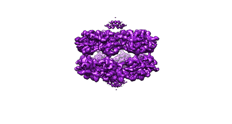 emd_3595.png emd_3595.png | 109.8 KB | ||
| Filedesc metadata | emd-3595.cif.gz | 5.6 KB | ||
| アーカイブディレクトリ |  http://ftp.pdbj.org/pub/emdb/structures/EMD-3595ftp://ftp.pdbj.org/pub/emdb/structures/EMD-3595 http://ftp.pdbj.org/pub/emdb/structures/EMD-3595ftp://ftp.pdbj.org/pub/emdb/structures/EMD-3595 | HTTPS FTP |
-関連構造データ


-リンク
| EMDBのページ | EMDB (EBI/PDBe) / EMDataResource |
|---|---|
| 「今月の分子」の関連する項目 |


-マップ
| ファイル | ダウンロード / ファイル: emd_3595.map.gz / 形式: CCP4 / 大きさ: 64 MB / タイプ: IMAGE STORED AS FLOATING POINT NUMBER (4 BYTES) | ||||||||||||||||||||||||||||||||||||||||||||||||||||||||||||
|---|---|---|---|---|---|---|---|---|---|---|---|---|---|---|---|---|---|---|---|---|---|---|---|---|---|---|---|---|---|---|---|---|---|---|---|---|---|---|---|---|---|---|---|---|---|---|---|---|---|---|---|---|---|---|---|---|---|---|---|---|---|
| 注釈 | Retinoschisin R141H D8 Hexadecameric Complex | ||||||||||||||||||||||||||||||||||||||||||||||||||||||||||||
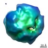
| 投影像・断面図 | 画像のコントロール
画像は Spider により作成 | ||||||||||||||||||||||||||||||||||||||||||||||||||||||||||||
| ボクセルのサイズ | X=Y=Z: 1.29 Å | ||||||||||||||||||||||||||||||||||||||||||||||||||||||||||||
| 密度 |
| ||||||||||||||||||||||||||||||||||||||||||||||||||||||||||||
| 対称性 | 空間群: 1 | ||||||||||||||||||||||||||||||||||||||||||||||||||||||||||||
| 詳細 | EMDB XML:
CCP4マップ ヘッダ情報:
| ||||||||||||||||||||||||||||||||||||||||||||||||||||||||||||
 Z (Sec.)
 Y (Row.)
 X (Col.)


-添付データ
- 試料の構成要素
-全体 : Retinoschisin
| 全体 | 名称: Retinoschisin |
|---|---|
| 要素 |
|
-超分子 #1: Retinoschisin
| 超分子 | 名称: Retinoschisin / タイプ: complex / ID: 1 / 親要素: 0 / 含まれる分子: all 詳細: Hexadecameric complex of sixteen retinoschisin molecules |
|---|---|
| 由来(天然) | 生物種: Homo sapiens (ヒト) |
-分子 #1: Retinoschisin
| 分子 | 名称: Retinoschisin / タイプ: protein_or_peptide / ID: 1 / コピー数: 16 / 光学異性体: LEVO |
|---|---|
| 由来(天然) | 生物種: Homo sapiens (ヒト) |
| 分子量 | 理論値: 23.041902 KDa |
| 組換発現 | 生物種: Homo sapiens (ヒト) |
| 配列 | 文字列: STEDEGEDPW YQKACKCDCQ GGPNALWSAG ATSLDCIPEC PYHKPLGFES GEVTPDQITC SNPEQYVGWY SSWTANKARL NSQGFGCAW LSKFQDSSQW LQIDLKEIKV ISGILTQGHC DIDEWMTKYS VQYRTDERLN WIYYKDQTGN NRVFYGNSDR T STVQNLLR ...文字列: STEDEGEDPW YQKACKCDCQ GGPNALWSAG ATSLDCIPEC PYHKPLGFES GEVTPDQITC SNPEQYVGWY SSWTANKARL NSQGFGCAW LSKFQDSSQW LQIDLKEIKV ISGILTQGHC DIDEWMTKYS VQYRTDERLN WIYYKDQTGN NRVFYGNSDR T STVQNLLR PPIISRFIRL IPLGWHVRIA IRMELLECVS KCA UniProtKB: Retinoschisin |
-実験情報
-構造解析
| 手法 | クライオ電子顕微鏡法 |
|---|---|
 解析 解析 | 単粒子再構成法 |
| 試料の集合状態 | particle |
-試料調製
| 濃度 | 0.1 mg/mL | |||||||||
|---|---|---|---|---|---|---|---|---|---|---|
| 緩衝液 | pH: 7.4 構成要素:
| |||||||||
| グリッド | モデル: C-flat-2/2 / 材質: COPPER / メッシュ: 400 / 前処理 - タイプ: GLOW DISCHARGE / 前処理 - 時間: 25 sec. | |||||||||
| 凍結 | 凍結剤: ETHANE / チャンバー内湿度: 100 % / チャンバー内温度: 277 K / 装置: FEI VITROBOT MARK I | |||||||||
| 詳細 | The sample was monodisperse and visible |
- 電子顕微鏡法
| 顕微鏡 | FEI TITAN KRIOS |
|---|---|
| 撮影 | フィルム・検出器のモデル: GATAN K2 SUMMIT (4k x 4k) 検出モード: COUNTING / デジタル化 - 画像ごとのフレーム数: 2-8 / 撮影したグリッド数: 1 / 実像数: 1200 / 平均露光時間: 0.5 sec. / 平均電子線量: 2.8 e/Å2 |
| 電子線 | 加速電圧: 300 kV / 電子線源:  FIELD EMISSION GUN FIELD EMISSION GUN |
| 電子光学系 | 照射モード: SPOT SCAN / 撮影モード: BRIGHT FIELD / Cs: 2.7 mm / 最大 デフォーカス(公称値): 4.5 µm / 最小 デフォーカス(公称値): 1.0 µm / 倍率(公称値): 105000 |
| 試料ステージ | 試料ホルダーモデル: FEI TITAN KRIOS AUTOGRID HOLDER ホルダー冷却材: NITROGEN |
| 実験機器 |  モデル: Titan Krios / 画像提供: FEI Company |
+画像解析

-原子モデル構築 1
| 精密化 | プロトコル: FLEXIBLE FIT 当てはまり具合の基準: Cross-correlation coefficient |
|---|---|
| 得られたモデル |  PDB-5n6w: |
