 Movie
Movie Controller

[English] 日本語
 Yorodumi
- EMDB-1279: Different quaternary structures of human RECQ1 are associated wit... -
+ Open data

- Basic information
| Entry | Database: EMDB / ID: EMD-1279 | |||||||||
|---|---|---|---|---|---|---|---|---|---|---|
| Title | Different quaternary structures of human RECQ1 are associated with its dual enzymatic activity. | |||||||||
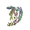
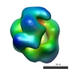
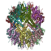
 Map data Map data | Map of K119R mutant RECQ1 bound to ssDNA, filtered using a 3D Gaussianlow-pass filter to a half-width of 17 Angstrom | |||||||||
 Sample Sample |
| |||||||||
| Biological species |  Homo sapiens (human) Homo sapiens (human) | |||||||||
| Method | single particle reconstruction / cryo EM / negative staining / Resolution: 24.0 Å | |||||||||
 Authors Authors | Muzzolini L / Beuron F / Patwardhan A / Popuri V / Cui S / Niccolini B / Rappas M / Freemont PS / Vindigni A | |||||||||
 Citation Citation | Journal: PLoS Biol / Year: 2007 Title: Different quaternary structures of human RECQ1 are associated with its dual enzymatic activity. Authors: Laura Muzzolini / Fabienne Beuron / Ardan Patwardhan / Venkateswarlu Popuri / Sheng Cui / Benedetta Niccolini / Mathieu Rappas / Paul S Freemont / Alessandro Vindigni /  Abstract: RecQ helicases are essential for the maintenance of chromosome stability. In addition to DNA unwinding, some RecQ enzymes have an intrinsic DNA strand annealing activity. The function of this dual ...RecQ helicases are essential for the maintenance of chromosome stability. In addition to DNA unwinding, some RecQ enzymes have an intrinsic DNA strand annealing activity. The function of this dual enzymatic activity and the mechanism that regulates it is, however, unknown. Here, we describe two quaternary forms of the human RECQ1 helicase, higher-order oligomers consistent with pentamers or hexamers, and smaller oligomers consistent with monomers or dimers. Size exclusion chromatography and transmission electron microscopy show that the equilibrium between the two assembly states is affected by single-stranded DNA (ssDNA) and ATP binding, where ATP or ATPgammaS favors the smaller oligomeric form. Our three-dimensional electron microscopy reconstructions of human RECQ1 reveal a complex cage-like structure of approximately 120 A x 130 A with a central pore. This oligomeric structure is stabilized under conditions in which RECQ1 is proficient in strand annealing. In contrast, competition experiments with the ATPase-deficient K119R and E220Q mutants indicate that RECQ1 monomers, or tight binding dimers, are required for DNA unwinding. Collectively, our findings suggest that higher-order oligomers are associated with DNA strand annealing, and lower-order oligomers with DNA unwinding. | |||||||||
| History |
|
- Structure visualization
| Movie |
 Movie viewer Movie viewer |
|---|---|
| Structure viewer | EM map: SurfViewMolmilJmol/JSmol |
| Supplemental images |

- Downloads & links
-EMDB archive
| Map data | emd_1279.map.gz | 3.6 MB | EMDB map data format | |
|---|---|---|---|---|
| Header (meta data) | emd-1279-v30.xmlemd-1279.xml | 9 KB 9 KB | Display Display | EMDB header |
| Images |  1279.gif 1279.gif | 62 KB | ||
| Archive directory |  http://ftp.pdbj.org/pub/emdb/structures/EMD-1279ftp://ftp.pdbj.org/pub/emdb/structures/EMD-1279 http://ftp.pdbj.org/pub/emdb/structures/EMD-1279ftp://ftp.pdbj.org/pub/emdb/structures/EMD-1279 | HTTPS FTP |
-Related structure data


-Links
| EMDB pages | EMDB (EBI/PDBe) / EMDataResource |
|---|
-Map
| File | Download / File: emd_1279.map.gz / Format: CCP4 / Size: 3.7 MB / Type: IMAGE STORED AS FLOATING POINT NUMBER (4 BYTES) | ||||||||||||||||||||||||||||||||||||||||||||||||||||||||||||||||||||
|---|---|---|---|---|---|---|---|---|---|---|---|---|---|---|---|---|---|---|---|---|---|---|---|---|---|---|---|---|---|---|---|---|---|---|---|---|---|---|---|---|---|---|---|---|---|---|---|---|---|---|---|---|---|---|---|---|---|---|---|---|---|---|---|---|---|---|---|---|---|
| Annotation | Map of K119R mutant RECQ1 bound to ssDNA, filtered using a 3D Gaussianlow-pass filter to a half-width of 17 Angstrom | ||||||||||||||||||||||||||||||||||||||||||||||||||||||||||||||||||||
| Projections & slices | Image control
Images are generated by Spider. | ||||||||||||||||||||||||||||||||||||||||||||||||||||||||||||||||||||
| Voxel size | X=Y=Z: 2.54 Å | ||||||||||||||||||||||||||||||||||||||||||||||||||||||||||||||||||||
| Density |
| ||||||||||||||||||||||||||||||||||||||||||||||||||||||||||||||||||||
| Symmetry | Space group: 1 | ||||||||||||||||||||||||||||||||||||||||||||||||||||||||||||||||||||
| Details | EMDB XML:
CCP4 map header:
| ||||||||||||||||||||||||||||||||||||||||||||||||||||||||||||||||||||
 Z (Sec.)
 Y (Row.)
 X (Col.)


-Supplemental data
- Sample components
-Entire : K119R mutant RECQ1 and ssDNA
| Entire | Name: K119R mutant RECQ1 and ssDNA |
|---|---|
| Components |
|
-Supramolecule #1000: K119R mutant RECQ1 and ssDNA
| Supramolecule | Name: K119R mutant RECQ1 and ssDNA / type: sample / ID: 1000 / Oligomeric state: Probably hexameric / Number unique components: 2 |
|---|---|
| Molecular weight | Experimental: 400 KDa / Theoretical: 440 KDa / Method: Size exclusion chromatography |
-Macromolecule #1: RECQ1
| Macromolecule | Name: RECQ1 / type: protein_or_peptide / ID: 1 / Name.synonym: RECQ1 / Number of copies: 6 / Oligomeric state: Possibly hexameric / Recombinant expression: Yes |
|---|---|
| Source (natural) | Organism: Homo sapiens (human) / synonym: Human |
| Molecular weight | Experimental: 400 KDa / Theoretical: 440 KDa |
-Macromolecule #2: Single stranded DNA - oligo-dT-30
| Macromolecule | Name: Single stranded DNA - oligo-dT-30 / type: dna / ID: 2 / Name.synonym: ssDNA / Classification: DNA / Structure: SINGLE STRANDED / Synthetic?: Yes |
|---|
-Experimental details
-Structure determination
| Method | negative staining, cryo EM |
|---|---|
 Processing Processing | single particle reconstruction |
| Aggregation state | particle |
-Sample preparation
| Concentration | 0.015 mg/mL |
|---|---|
| Buffer | pH: 7.4 / Details: 20mM TrisHCl, 150 mM KCl, 2 mM MgCl2 |
| Staining | Type: NEGATIVE Details: RECQ1 was incubated with a 1.5 molar excess of ssDNA, adsorbed onto a glow-discharged carbon-coated grid and negatively-stained with 1% uranyl acetate |
| Vitrification | Cryogen name: ETHANE |
- Electron microscopy
| Microscope | FEI/PHILIPS CM200FEG |
|---|---|
| Date | Jun 6, 2005 |
| Image recording | Category: CCD / Film or detector model: KODAK SO-163 FILM / Digitization - Sampling interval: 6.35 µm / Bits/pixel: 8 |
| Electron beam | Acceleration voltage: 200 kV / Electron source:  FIELD EMISSION GUN FIELD EMISSION GUN |
| Electron optics | Calibrated magnification: 48600 / Illumination mode: FLOOD BEAM / Imaging mode: BRIGHT FIELD / Nominal magnification: 50000 |
| Sample stage | Specimen holder: Eucentric / Specimen holder model: GATAN LIQUID NITROGEN |
-Image processing
| Final reconstruction | Applied symmetry - Point group: C1 (asymmetric) / Algorithm: OTHER / Resolution.type: BY AUTHOR / Resolution: 24.0 Å / Resolution method: FSC 0.5 CUT-OFF / Software - Name: Imagic / Number images used: 1163 |
|---|---|
| Final angle assignment | Details: Imagic |
| Final two d classification | Number classes: 87 |
