 Movie
Movie Controller


+ Open data

- Basic information
| Entry | Database: PDB / ID: 4hvz | ||||||
|---|---|---|---|---|---|---|---|
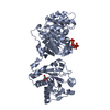
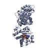
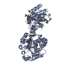
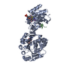
| Title | Crystal structure of brucella abortus immunogenic BP26 protein | ||||||
 Components Components | 26 kDa periplasmic immunogenic protein | ||||||
 Keywords Keywords | MEMBRANE PROTEIN / multimerization / SIMPL domain / infection | ||||||
| Function / homology |  Function and homology information Function and homology information | ||||||
| Biological species |  Brucella abortus (bacteria) Brucella abortus (bacteria) | ||||||
| Method |  X-RAY DIFFRACTION / SYNCHROTRON / SAD / Resolution: 3.5 Å X-RAY DIFFRACTION / SYNCHROTRON / SAD / Resolution: 3.5 Å | ||||||
 Authors Authors | Kim, D. / Park, J. / Oh, B. / Song, J. | ||||||
 Citation Citation | Journal: J.Mol.Biol. / Year: 2013 Title: Brucella Immunogenic BP26 Forms a Channel-like Structure. Authors: Kim, D. / Park, J. / Kim, S.J. / Soh, Y.M. / Kim, H.M. / Oh, B.H. / Song, J.J. | ||||||
| History |
|
- Structure visualization
| Structure viewer | Molecule: MolmilJmol/JSmol |
|---|
- Downloads & links
-Download
| PDBx/mmCIF format | 4hvz.cif.gz | 165.3 KB | Display | PDBx/mmCIF format |
|---|---|---|---|---|
| PDB format | pdb4hvz.ent.gz | 134.6 KB | Display | PDB format |
| PDBx/mmJSON format | 4hvz.json.gz | Tree view | PDBx/mmJSON format | |
| Others |  Other downloads Other downloads |
-Validation report
| Arichive directory | https://data.pdbj.org/pub/pdb/validation_reports/hv/4hvzftp://data.pdbj.org/pub/pdb/validation_reports/hv/4hvz | HTTPS FTP |
|---|
-Related structure data
| Similar structure data |
|---|


-Links
 PDBj
PDBj- Assembly
| Deposited unit | 
| ||||||||
|---|---|---|---|---|---|---|---|---|---|
| 1 | 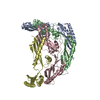
| ||||||||
| Unit cell |
|
-Components
| #1: Protein | Mass: 23802.096 Da / Num. of mol.: 4 / Fragment: BP26 Source method: isolated from a genetically manipulated source Source: (gene. exp.) Brucella abortus (bacteria) / Strain: S19 / Gene: bp26, BAbS19_I13950 / Plasmid: pET28a / Production host: |
|---|
-Experimental details
-Experiment
| Experiment | Method: X-RAY DIFFRACTION / Number of used crystals: 1 |
|---|
- Sample preparation
| Crystal | Density Matthews: 5.61 Å3/Da / Density % sol: 78.08 % |
|---|---|
| Crystal grow | Temperature: 293 K / Method: vapor diffusion, hanging drop / pH: 8.5 Details: 2.4M AMS, 100mM NaCl, pH 8.5, VAPOR DIFFUSION, HANGING DROP, temperature 293K |
-Data collection
| Diffraction | Mean temperature: 110 K |
|---|---|
| Diffraction source | Source: SYNCHROTRON / Type: OTHER / Wavelength: 0.97941 Å |
| Detector | Type: ADSC QUANTUM 315r / Detector: CCD / Date: Jun 24, 2012 |
| Radiation | Protocol: SINGLE WAVELENGTH / Monochromatic (M) / Laue (L): M / Scattering type: x-ray |
| Radiation wavelength | Wavelength: 0.97941 Å / Relative weight: 1 |
| Reflection | Resolution: 3.5→30 Å / Num. obs: 26996 / Observed criterion σ(F): 0 / Redundancy: 5.9 % / Rsym value: 0.08 / Net I/σ(I): 31.9 |
| Reflection shell | Resolution: 3.5→3.56 Å / Redundancy: 6.1 % / Rmerge(I) obs: 0.531 / Mean I/σ(I) obs: 3.8 / % possible all: 99.3 |
- Processing
| Software |
| ||||||||||||||||||||||||||||||||||||||||||||||||||||||||||||||||||||||||||||||||
|---|---|---|---|---|---|---|---|---|---|---|---|---|---|---|---|---|---|---|---|---|---|---|---|---|---|---|---|---|---|---|---|---|---|---|---|---|---|---|---|---|---|---|---|---|---|---|---|---|---|---|---|---|---|---|---|---|---|---|---|---|---|---|---|---|---|---|---|---|---|---|---|---|---|---|---|---|---|---|---|---|---|
| Refinement | Method to determine structure: SAD / Resolution: 3.5→29.96 Å / Rfactor Rfree error: 0.006 / Data cutoff high absF: 101166.96 / Data cutoff low absF: 0 / Isotropic thermal model: RESTRAINED / Cross valid method: THROUGHOUT / σ(F): 0 / Details: BULK SOLVENT MODEL USED
| ||||||||||||||||||||||||||||||||||||||||||||||||||||||||||||||||||||||||||||||||
| Solvent computation | Solvent model: FLAT MODEL / Bsol: 71.6865 Å2 / ksol: 0.3 e/Å3 | ||||||||||||||||||||||||||||||||||||||||||||||||||||||||||||||||||||||||||||||||
| Displacement parameters | Biso mean: 121.3 Å2
| ||||||||||||||||||||||||||||||||||||||||||||||||||||||||||||||||||||||||||||||||
| Refine analyze |
| ||||||||||||||||||||||||||||||||||||||||||||||||||||||||||||||||||||||||||||||||
| Refinement step | Cycle: LAST / Resolution: 3.5→29.96 Å
| ||||||||||||||||||||||||||||||||||||||||||||||||||||||||||||||||||||||||||||||||
| Refine LS restraints |
| ||||||||||||||||||||||||||||||||||||||||||||||||||||||||||||||||||||||||||||||||
| LS refinement shell | Resolution: 3.5→3.72 Å / Rfactor Rfree error: 0.02 / Total num. of bins used: 6
| ||||||||||||||||||||||||||||||||||||||||||||||||||||||||||||||||||||||||||||||||
| Xplor file | Serial no: 1 / Param file: protein_rep.param / Topol file: protein.top |
