 Movie
Movie Controller


+ Open data

- Basic information
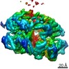
| Entry | Database: EMDB / ID: EMD-3207 | |||||||||
|---|---|---|---|---|---|---|---|---|---|---|
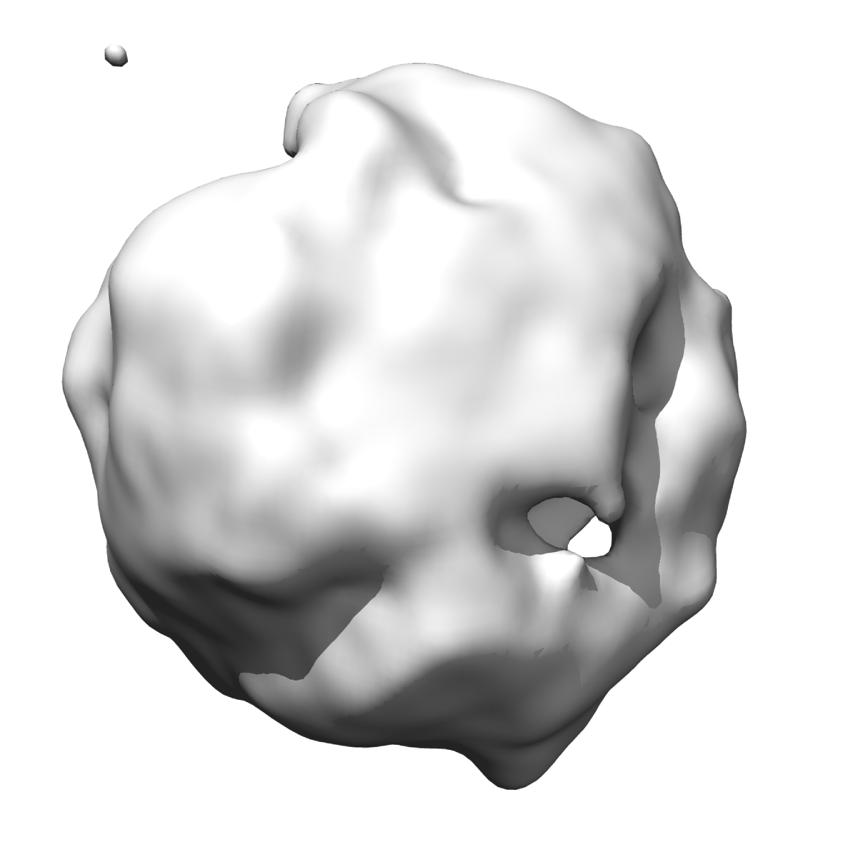
| Title | Negative stain structure of the Rix1-Ipi1dN50-Ipi3 complex | |||||||||
 Map data Map data | Reconstruction of mutant Rix1-Ipi1dN50-Ipi3 complex | |||||||||
 Sample Sample |
| |||||||||
 Keywords Keywords | ribosome / ribosome biogenesis / ribosome assembly / pre-60S / 5S RNP / assembly intermediate / negative-stain / Rix1 complex / Rix1 / Ipi1 / Ipi3 | |||||||||
| Biological species |  | |||||||||
| Method | single particle reconstruction / negative staining / Resolution: 18.0 Å | |||||||||
 Authors Authors | Barrio-Garcia C / Thoms M / Flemming D / Kater L / Berninghausen O / Bassler J / Beckmann R / Hurt E | |||||||||
 Citation Citation | Journal: Nat Struct Mol Biol / Year: 2016 Title: Architecture of the Rix1-Rea1 checkpoint machinery during pre-60S-ribosome remodeling. Authors: Clara Barrio-Garcia / Matthias Thoms / Dirk Flemming / Lukas Kater / Otto Berninghausen / Jochen Baßler / Roland Beckmann / Ed Hurt /  Abstract: Ribosome synthesis is catalyzed by ∼200 assembly factors, which facilitate efficient production of mature ribosomes. Here, we determined the cryo-EM structure of a Saccharomyces cerevisiae ...Ribosome synthesis is catalyzed by ∼200 assembly factors, which facilitate efficient production of mature ribosomes. Here, we determined the cryo-EM structure of a Saccharomyces cerevisiae nucleoplasmic pre-60S particle containing the dynein-related 550-kDa Rea1 AAA(+) ATPase and the Rix1 subcomplex. This particle differs from its preceding state, the early Arx1 particle, by two massive structural rearrangements: an ∼180° rotation of the 5S ribonucleoprotein complex and the central protuberance (CP) rRNA helices, and the removal of the 'foot' structure from the 3' end of the 5.8S rRNA. Progression from the Arx1 to the Rix1 particle was blocked by mutational perturbation of the Rix1-Rea1 interaction but not by a dominant-lethal Rea1 AAA(+) ATPase-ring mutant. After remodeling, the Rix1 subcomplex and Rea1 become suitably positioned to sense correct structural maturation of the CP, which allows unidirectional progression toward mature ribosomes. | |||||||||
| History |
|
- Structure visualization
| Movie |
 Movie viewer Movie viewer |
|---|---|
| Structure viewer | EM map: SurfViewMolmilJmol/JSmol |
| Supplemental images |
- Downloads & links
-EMDB archive
| Map data | emd_3207.map.gz | 6.3 MB | EMDB map data format | |
|---|---|---|---|---|
| Header (meta data) | emd-3207-v30.xmlemd-3207.xml | 9.7 KB 9.7 KB | Display Display | EMDB header |
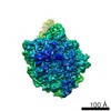
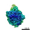
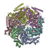
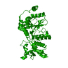
| Images |  EMD-3207_Rix1-subcomplex.png EMD-3207_Rix1-subcomplex.png | 393.3 KB | ||
| Archive directory |  http://ftp.pdbj.org/pub/emdb/structures/EMD-3207ftp://ftp.pdbj.org/pub/emdb/structures/EMD-3207 http://ftp.pdbj.org/pub/emdb/structures/EMD-3207ftp://ftp.pdbj.org/pub/emdb/structures/EMD-3207 | HTTPS FTP |
-Related structure data


-Links
| EMDB pages | EMDB (EBI/PDBe) / EMDataResource |
|---|---|
| Related items in Molecule of the Month |

-Map
| File | Download / File: emd_3207.map.gz / Format: CCP4 / Size: 21.7 MB / Type: IMAGE STORED AS FLOATING POINT NUMBER (4 BYTES) | ||||||||||||||||||||||||||||||||||||||||||||||||||||||||||||||||||||
|---|---|---|---|---|---|---|---|---|---|---|---|---|---|---|---|---|---|---|---|---|---|---|---|---|---|---|---|---|---|---|---|---|---|---|---|---|---|---|---|---|---|---|---|---|---|---|---|---|---|---|---|---|---|---|---|---|---|---|---|---|---|---|---|---|---|---|---|---|---|
| Annotation | Reconstruction of mutant Rix1-Ipi1dN50-Ipi3 complex | ||||||||||||||||||||||||||||||||||||||||||||||||||||||||||||||||||||
| Projections & slices | Image control
Images are generated by Spider. | ||||||||||||||||||||||||||||||||||||||||||||||||||||||||||||||||||||
| Voxel size | X=Y=Z: 1.65 Å | ||||||||||||||||||||||||||||||||||||||||||||||||||||||||||||||||||||
| Density |
| ||||||||||||||||||||||||||||||||||||||||||||||||||||||||||||||||||||
| Symmetry | Space group: 1 | ||||||||||||||||||||||||||||||||||||||||||||||||||||||||||||||||||||
| Details | EMDB XML:
CCP4 map header:
| ||||||||||||||||||||||||||||||||||||||||||||||||||||||||||||||||||||
 Z (Sec.)
 Y (Row.)
 X (Col.)


-Supplemental data
- Sample components
-Entire : Rix1-Ipi1dN50-Ipi3 complex
| Entire | Name: Rix1-Ipi1dN50-Ipi3 complex |
|---|---|
| Components |
|
-Supramolecule #1000: Rix1-Ipi1dN50-Ipi3 complex
| Supramolecule | Name: Rix1-Ipi1dN50-Ipi3 complex / type: sample / ID: 1000 Oligomeric state: 2 copies of Rix1, 1 copy of Ipi1 and 2 copies of Ipi3 Number unique components: 3 |
|---|---|
| Molecular weight | Experimental: 330 KDa / Theoretical: 334 KDa Method: size-exclusion chromatography coupled to multi-angle light scattering (SEC-MALS) |
-Macromolecule #1: Rix1
| Macromolecule | Name: Rix1 / type: protein_or_peptide / ID: 1 / Name.synonym: Rix1 complex / Number of copies: 2 / Oligomeric state: heterotrimer / Recombinant expression: No |
|---|---|
| Source (natural) | Organism: |
| Molecular weight | Theoretical: 867.5 KDa |
-Macromolecule #2: Ipi3
| Macromolecule | Name: Ipi3 / type: protein_or_peptide / ID: 2 / Number of copies: 2 / Oligomeric state: heterotrimer / Recombinant expression: No |
|---|---|
| Source (natural) | Organism: |
| Molecular weight | Experimental: 61.78 KDa |
-Macromolecule #3: Ipi1dN50
| Macromolecule | Name: Ipi1dN50 / type: protein_or_peptide / ID: 3 / Number of copies: 1 / Oligomeric state: heterotrimer / Recombinant expression: No |
|---|---|
| Source (natural) | Organism: |
| Molecular weight | Theoretical: 37.87 KDa |
-Experimental details
-Structure determination
| Method | negative staining |
|---|---|
 Processing Processing | single particle reconstruction |
| Aggregation state | particle |
-Sample preparation
| Buffer | pH: 7.5 Details: 20 mM Hepes, 200 mM NaCl, 10 mM KCl, 10 mM MgCl2 and 1 mM DTT |
|---|---|
| Staining | Type: NEGATIVE / Details: stained with 2% (w/v) uranyl acetate |
| Vitrification | Cryogen name: NONE / Instrument: OTHER |
- Electron microscopy
| Microscope | FEI TECNAI F20 |
|---|---|
| Date | Mar 6, 2013 |
| Image recording | Category: CCD / Film or detector model: FEI EAGLE (4k x 4k) / Number real images: 25 |
| Electron beam | Acceleration voltage: 200 kV / Electron source:  FIELD EMISSION GUN FIELD EMISSION GUN |
| Electron optics | Illumination mode: SPOT SCAN / Imaging mode: BRIGHT FIELD / Cs: 1.2 mm / Nominal magnification: 62000 |
| Sample stage | Specimen holder model: SIDE ENTRY, EUCENTRIC |
| Experimental equipment |  Model: Tecnai F20 / Image courtesy: FEI Company |
-Image processing
| Details | The particles were selected using the interactive selection program BOXER |
|---|---|
| Final reconstruction | Applied symmetry - Point group: C1 (asymmetric) / Resolution.type: BY AUTHOR / Resolution: 18.0 Å / Resolution method: OTHER / Software - Name: IMAGIC-4D, BOXER / Number images used: 4267 |
