 Movie
Movie Controller

[English] 日本語
 Yorodumi
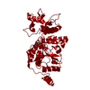
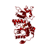
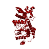
- PDB-1l9v: Non Structural protein encoded by gene segment 8 of rotavirus (NS... -
+ Open data

- Basic information
| Entry | Database: PDB / ID: 1l9v | ||||||
|---|---|---|---|---|---|---|---|
| Title | Non Structural protein encoded by gene segment 8 of rotavirus (NSP2), an NTPase, ssRNA binding and nucleic acid helix-destabilizing protein | ||||||
 Components Components | Rotavirus-NSP2 | ||||||
 Keywords Keywords | VIRAL PROTEIN / alpha/beta protein / HIT-like fold / Octamer / Two domain protein | ||||||
| Function / homology |  Function and homology information Function and homology informationnucleoside diphosphate kinase activity / viral genome replication / Hydrolases; Acting on acid anhydrides; Acting on acid anhydrides to facilitate cellular and subcellular movement / ribonucleoside triphosphate phosphatase activity / host cell cytoplasm / RNA binding / ATP binding / metal ion binding Similarity search - Function | ||||||
| Biological species |   Simian 11 rotavirus Simian 11 rotavirus | ||||||
| Method |  X-RAY DIFFRACTION / SYNCHROTRON / MAD / Resolution: 2.6 Å X-RAY DIFFRACTION / SYNCHROTRON / MAD / Resolution: 2.6 Å | ||||||
 Authors Authors | Jayaram, H. / Taraporewala, Z. / Patton, J.T. / Prasad, B.V. | ||||||
 Citation Citation | Journal: Nature / Year: 2002 Title: Rotavirus protein involved in genome replication and packaging exhibits a HIT-like fold. Authors: Jayaram, H. / Taraporewala, Z. / Patton, J.T. / Prasad, B.V. | ||||||
| History |
|
- Structure visualization
| Structure viewer | Molecule: MolmilJmol/JSmol |
|---|
- Downloads & links
-Download
| PDBx/mmCIF format | 1l9v.cif.gz | 67.6 KB | Display | PDBx/mmCIF format |
|---|---|---|---|---|
| PDB format | pdb1l9v.ent.gz | 52.1 KB | Display | PDB format |
| PDBx/mmJSON format | 1l9v.json.gz | Tree view | PDBx/mmJSON format | |
| Others |  Other downloads Other downloads |
-Validation report
| Arichive directory | https://data.pdbj.org/pub/pdb/validation_reports/l9/1l9vftp://data.pdbj.org/pub/pdb/validation_reports/l9/1l9v | HTTPS FTP |
|---|
-Related structure data
| Similar structure data |
|---|


-Links
 PDBj
PDBj- Assembly
| Deposited unit | 
| ||||||||
|---|---|---|---|---|---|---|---|---|---|
| 1 | x 8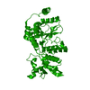
| ||||||||
| Unit cell |
| ||||||||
| Details | Octamer - obtained by applying I422 symmtery |
-Components
| #1: Protein | Mass: 36618.258 Da / Num. of mol.: 1 / Fragment: NSP2 of Rotavirus Source method: isolated from a genetically manipulated source Source: (gene. exp.) Simian 11 rotavirus (serotype 3 / strain SA11-Ramig)Species: Rotavirus A / Strain: SA11-Ramig / Gene: gene segment 8 / Plasmid: pQe60 (Qiagen) / Production host:  |
|---|---|
| #2: Water | ChemComp-HOH /  Mass: 18.015 Da / Num. of mol.: 1 / Source method: isolated from a natural source / Formula: H2O Mass: 18.015 Da / Num. of mol.: 1 / Source method: isolated from a natural source / Formula: H2O |
-Experimental details
-Experiment
| Experiment | Method: X-RAY DIFFRACTION / Number of used crystals: 1 |
|---|
- Sample preparation
| Crystal | Density Matthews: 3.07 Å3/Da / Density % sol: 59.9 % | ||||||||||||||||||||||||||||||
|---|---|---|---|---|---|---|---|---|---|---|---|---|---|---|---|---|---|---|---|---|---|---|---|---|---|---|---|---|---|---|---|
| Crystal grow | Temperature: 298 K / Method: vapor diffusion, hanging drop / pH: 7.25 Details: 15% Peg 6000, 0.2 M Magnesium acetate, 0.1M Tris-HCl. Protein buffer 0.2M MgCl2, 0.5 mM EDTA , 0.5 mM DTT, 2mM Tris-HCl pH7.2. 0.2 M MgCl2 was necessary to make protein monodisperse. Protein: ...Details: 15% Peg 6000, 0.2 M Magnesium acetate, 0.1M Tris-HCl. Protein buffer 0.2M MgCl2, 0.5 mM EDTA , 0.5 mM DTT, 2mM Tris-HCl pH7.2. 0.2 M MgCl2 was necessary to make protein monodisperse. Protein:precipitant ratio 1:1. Plate like crystals (0.08 mm X 0.2 mm X 0.2 mm)Crystals grew in two to three weeks, pH 7.25, VAPOR DIFFUSION, HANGING DROP, temperature 298K | ||||||||||||||||||||||||||||||
| Crystal grow | *PLUS PH range low: 7.5 / PH range high: 6.5 | ||||||||||||||||||||||||||||||
| Components of the solutions | *PLUS
|
-Data collection
| Diffraction |
| |||||||||
|---|---|---|---|---|---|---|---|---|---|---|
| Diffraction source | Source: SYNCHROTRON / Site: NSLS  / Beamline: X8C / Wavelength: 0.9795 Å / Beamline: X8C / Wavelength: 0.9795 Å | |||||||||
| Detector | Type: ADSC QUANTUM 4 / Detector: CCD / Date: Feb 24, 2001 / Details: Mirrors | |||||||||
| Radiation | Monochromator: Two crystal monochromator (non-dispersive geometry) with Si(111) crystals Protocol: MAD / Monochromatic (M) / Laue (L): M / Scattering type: x-ray | |||||||||
| Radiation wavelength | Wavelength: 0.9795 Å / Relative weight: 1 | |||||||||
| Reflection | Resolution: 2.6→40 Å / Num. all: 14403 / Num. obs: 75176 / % possible obs: 92.5 % / Redundancy: 5.2 % / Biso Wilson estimate: 46.5 Å2 / Rmerge(I) obs: 0.068 / Net I/σ(I): 10.1 | |||||||||
| Reflection shell | Resolution: 2.6→2.69 Å / % possible all: 94.2 | |||||||||
| Reflection | *PLUS Highest resolution: 2.6 Å / Lowest resolution: 40 Å / % possible obs: 95.9 % | |||||||||
| Reflection shell | *PLUS % possible obs: 94.2 % / Rmerge(I) obs: 0.314 / Mean I/σ(I) obs: 2.8 |
- Processing
| Software |
| ||||||||||||||||||||||||||||||||||||||||||||||||||||||||||||||||||||||||||||||||
|---|---|---|---|---|---|---|---|---|---|---|---|---|---|---|---|---|---|---|---|---|---|---|---|---|---|---|---|---|---|---|---|---|---|---|---|---|---|---|---|---|---|---|---|---|---|---|---|---|---|---|---|---|---|---|---|---|---|---|---|---|---|---|---|---|---|---|---|---|---|---|---|---|---|---|---|---|---|---|---|---|---|
| Refinement | Method to determine structure: MAD / Resolution: 2.6→38.34 Å / Rfactor Rfree error: 0.008 / Data cutoff high absF: 196804.06 / Data cutoff low absF: 0 / Isotropic thermal model: RESTRAINED / Cross valid method: THROUGHOUT / σ(F): 0 / Stereochemistry target values: CNS_PARAM Files
| ||||||||||||||||||||||||||||||||||||||||||||||||||||||||||||||||||||||||||||||||
| Solvent computation | Solvent model: FLAT MODEL / Bsol: 45.3326 Å2 / ksol: 0.357545 e/Å3 | ||||||||||||||||||||||||||||||||||||||||||||||||||||||||||||||||||||||||||||||||
| Displacement parameters | Biso mean: 54.5 Å2
| ||||||||||||||||||||||||||||||||||||||||||||||||||||||||||||||||||||||||||||||||
| Refine analyze |
| ||||||||||||||||||||||||||||||||||||||||||||||||||||||||||||||||||||||||||||||||
| Refinement step | Cycle: LAST / Resolution: 2.6→38.34 Å
| ||||||||||||||||||||||||||||||||||||||||||||||||||||||||||||||||||||||||||||||||
| Refine LS restraints |
| ||||||||||||||||||||||||||||||||||||||||||||||||||||||||||||||||||||||||||||||||
| LS refinement shell | Resolution: 2.6→2.76 Å / Rfactor Rfree error: 0.027 / Total num. of bins used: 6
| ||||||||||||||||||||||||||||||||||||||||||||||||||||||||||||||||||||||||||||||||
| Xplor file |
| ||||||||||||||||||||||||||||||||||||||||||||||||||||||||||||||||||||||||||||||||
| Refinement | *PLUS % reflection Rfree: 10 % / Rfactor obs: 0.24 / Rfactor Rfree: 0.2884 / Rfactor Rwork: 0.24 / Highest resolution: 2.6 Å / Lowest resolution: 40 Å | ||||||||||||||||||||||||||||||||||||||||||||||||||||||||||||||||||||||||||||||||
| Solvent computation | *PLUS | ||||||||||||||||||||||||||||||||||||||||||||||||||||||||||||||||||||||||||||||||
| Displacement parameters | *PLUS | ||||||||||||||||||||||||||||||||||||||||||||||||||||||||||||||||||||||||||||||||
| Refine LS restraints | *PLUS
| ||||||||||||||||||||||||||||||||||||||||||||||||||||||||||||||||||||||||||||||||
| LS refinement shell | *PLUS Rfactor obs: 0.335 |
